 Movie
Movie Controller

[English] 日本語
 Yorodumi
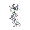
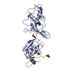
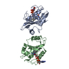
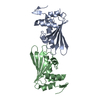
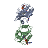
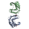
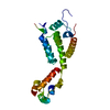
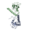
- PDB-1i3j: CRYSTAL STRUCTURE OF THE DNA-BINDING DOMAIN OF INTRON ENDONUCLEAS... -
+ Open data

- Basic information
| Entry | Database: PDB / ID: 1i3j | ||||||
|---|---|---|---|---|---|---|---|
| Title | CRYSTAL STRUCTURE OF THE DNA-BINDING DOMAIN OF INTRON ENDONUCLEASE I-TEVI WITH ITS SUBSTRATE | ||||||
 Components Components |
| ||||||
 Keywords Keywords | HYDROLASE/DNA /  PROTEIN-DNA COMPLEX / EXTENDED STRUCTURE / ZN-FINGER / MINOR GROOVE HELIX / HELIX-TURN-HELIX / HYDROLASE-DNA COMPLEX PROTEIN-DNA COMPLEX / EXTENDED STRUCTURE / ZN-FINGER / MINOR GROOVE HELIX / HELIX-TURN-HELIX / HYDROLASE-DNA COMPLEX | ||||||
| Function / homology |  Function and homology information Function and homology informationdouble-stranded DNA endonuclease activity / nucleic acid metabolic process / intron homing / DNA-binding transcription repressor activity / transcription repressor complex / endonuclease activity / sequence-specific DNA binding / Hydrolases; Acting on ester bonds / negative regulation of DNA-templated transcription / DNA binding / zinc ion bindingSimilarity search - Function | ||||||
| Biological species |  Enterobacteria phage T4 (virus) Enterobacteria phage T4 (virus) | ||||||
| Method | X-RAY DIFFRACTION / SYNCHROTRON / SIRAS / Resolution: 2.2 Å | ||||||
 Authors Authors | Van Roey, P. / Waddling, C.A. / Fox, K.M. / Belfort, M. / Derbyshire, V. | ||||||
 Citation Citation | Journal: EMBO J. / Year: 2001 Title: Intertwined structure of the DNA-binding domain of intron endonuclease I-TevI with its substrate. Authors: Van Roey, P. / Waddling, C.A. / Fox, K.M. / Belfort, M. / Derbyshire, V. | ||||||
| History |
|
- Structure visualization
| Structure viewer | Molecule: MolmilJmol/JSmol |
|---|
- Downloads & links
-Download
| PDBx/mmCIF format | 1i3j.cif.gz | 62.6 KB | Display | PDBx/mmCIF format |
|---|---|---|---|---|
| PDB format | pdb1i3j.ent.gz | 41.9 KB | Display | PDB format |
| PDBx/mmJSON format | 1i3j.json.gz | Tree view | PDBx/mmJSON format | |
| Others |  Other downloads Other downloads |
-Validation report
| Arichive directory | https://data.pdbj.org/pub/pdb/validation_reports/i3/1i3jftp://data.pdbj.org/pub/pdb/validation_reports/i3/1i3j | HTTPS FTP |
|---|
-Related structure data
| Similar structure data |
|---|


-Links
 PDBj
PDBj


- Assembly
| Deposited unit | 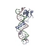
| ||||||||
|---|---|---|---|---|---|---|---|---|---|
| 1 |
| ||||||||
| Unit cell |
|
-Components
| #1: DNA chain | Mass: 6410.141 Da / Num. of mol.: 1 / Source method: obtained synthetically |
|---|---|
| #2: DNA chain | Mass: 6473.239 Da / Num. of mol.: 1 / Source method: obtained synthetically |
| #3: Protein | Mass: 13242.219 Da / Num. of mol.: 1 / Fragment: DNA-BINDING DOMAIN Source method: isolated from a genetically manipulated source Source: (gene. exp.) Enterobacteria phage T4 (virus) / Genus: T4-like viruses / Species: Enterobacteria phage T4 sensu lato / Plasmid: PET-3A / Production host:  Escherichia coli (E. coli) Escherichia coli (E. coli)References: UniProt: P13299, Hydrolases; Acting on ester bonds |
| #4: Chemical | ChemComp-ZN /   Mass: 65.409 Da / Num. of mol.: 1 / Source method: obtained synthetically / Formula: Zn Mass: 65.409 Da / Num. of mol.: 1 / Source method: obtained synthetically / Formula: Zn |
| #5: Water | ChemComp-HOH / Water Mass: 18.015 Da / Num. of mol.: 185 / Source method: isolated from a natural source / Formula: H2O Mass: 18.015 Da / Num. of mol.: 185 / Source method: isolated from a natural source / Formula: H2O |
-Experimental details
-Experiment
| Experiment | Method: X-RAY DIFFRACTION / Number of used crystals: 2 |
|---|
- Sample preparation
| Crystal | Density Matthews: 2.95 Å3/Da / Density % sol: 58.28 % | |||||||||||||||||||||||||
|---|---|---|---|---|---|---|---|---|---|---|---|---|---|---|---|---|---|---|---|---|---|---|---|---|---|---|
| Crystal grow | Temperature: 283 K / Method: vapor diffusion, hanging drop / pH: 6.5 Details: PEG3350, MES, glycerol, pH 6.5, VAPOR DIFFUSION, HANGING DROP, temperature 283K | |||||||||||||||||||||||||
| Components of the solutions |
| |||||||||||||||||||||||||
| Crystal grow | *PLUS Temperature: 10 ℃ | |||||||||||||||||||||||||
| Components of the solutions | *PLUS
|
-Data collection
| Diffraction |
| |||||||||||||||
|---|---|---|---|---|---|---|---|---|---|---|---|---|---|---|---|---|
| Diffraction source |
| |||||||||||||||
| Detector |
| |||||||||||||||
| Radiation | Protocol: SINGLE WAVELENGTH / Monochromatic (M) / Laue (L): M / Scattering type: x-ray | |||||||||||||||
| Radiation wavelength |
| |||||||||||||||
| Reflection | Resolution: 2.2→30 Å / Num. all: 88623 / Num. obs: 17561 / % possible obs: 99.4 % / Observed criterion σ(F): 0 / Observed criterion σ(I): 0 / Redundancy: 5.02 % / Rmerge(I) obs: 0.059 / Net I/σ(I): 9 | |||||||||||||||
| Reflection shell | Resolution: 2.2→2.28 Å / Redundancy: 4.9 % / Rmerge(I) obs: 0.389 / Mean I/σ(I) obs: 3.8 / % possible all: 98.8 | |||||||||||||||
| Reflection | *PLUS Rmerge(I) obs: 0.043 | |||||||||||||||
| Reflection shell | *PLUS % possible obs: 96.5 % |

- Processing
| Software |
| |||||||||||||||||||||||||
|---|---|---|---|---|---|---|---|---|---|---|---|---|---|---|---|---|---|---|---|---|---|---|---|---|---|---|
| Refinement | Method to determine structure: SIRAS / Resolution: 2.2→20 Å / Cross valid method: THROUGHOUT / σ(F): 0 / σ(I): 0 / Stereochemistry target values: Engh & Huber
| |||||||||||||||||||||||||
| Displacement parameters | Biso mean: 35.3 Å2 | |||||||||||||||||||||||||
| Refinement step | Cycle: LAST / Resolution: 2.2→20 Å
| |||||||||||||||||||||||||
| Refine LS restraints |
| |||||||||||||||||||||||||
| Software | *PLUS Name: CNS / Version: 0.9 / Classification: refinement | |||||||||||||||||||||||||
| Refinement | *PLUS Highest resolution: 2.2 Å / Lowest resolution: 20 Å / σ(F): 0 / Rfactor obs: 0.215 | |||||||||||||||||||||||||
| Solvent computation | *PLUS | |||||||||||||||||||||||||
| Displacement parameters | *PLUS Biso mean: 35.3 Å2 | |||||||||||||||||||||||||
| Refine LS restraints | *PLUS
|
