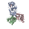
 Movie
Movie Controller


+ Open data

- Basic information
| Entry | Database: PDB / ID: 1hz4 | ||||||
|---|---|---|---|---|---|---|---|
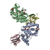
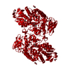
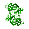
| Title | CRYSTAL STRUCTURE OF TRANSCRIPTION FACTOR MALT DOMAIN III | ||||||
 Components Components | MALT REGULATORY PROTEIN | ||||||
 Keywords Keywords |  TRANSCRIPTION ACTIVATOR / two-helix bundles / helix repeats / protein superhelix TRANSCRIPTION ACTIVATOR / two-helix bundles / helix repeats / protein superhelix | ||||||
| Function / homology |  Function and homology informationtrisaccharide binding / positive regulation of carbohydrate metabolic process / carbohydrate metabolic process / DNA-binding transcription factor activity / positive regulation of DNA-templated transcription / DNA binding / ATP binding / identical protein binding Function and homology informationtrisaccharide binding / positive regulation of carbohydrate metabolic process / carbohydrate metabolic process / DNA-binding transcription factor activity / positive regulation of DNA-templated transcription / DNA binding / ATP binding / identical protein bindingSimilarity search - Function | ||||||
| Biological species |  Escherichia coli (E. coli) Escherichia coli (E. coli) | ||||||
| Method | X-RAY DIFFRACTION / SYNCHROTRON / Resolution: 1.45 Å | ||||||
 Authors Authors | Steegborn, C. / Danot, O. / Clausen, T. / Huber, R. | ||||||
 Citation Citation | Journal: Structure / Year: 2001 Title: Crystal structure of transcription factor MalT domain III: a novel helix repeat fold implicated in regulated oligomerization. Authors: Steegborn, C. / Danot, O. / Huber, R. / Clausen, T. | ||||||
| History |
| ||||||
| Remark 300 | BIOMOLECULE: 1 THIS ENTRY CONTAINS THE CRYSTALLOGRAPHIC ASYMMETRIC UNIT WHICH CONSISTS OF 1 ... BIOMOLECULE: 1 THIS ENTRY CONTAINS THE CRYSTALLOGRAPHIC ASYMMETRIC UNIT WHICH CONSISTS OF 1 CHAIN(S). SEE REMARK 350 FOR INFORMATION ON GENERATING THE BIOLOGICAL MOLECULE(S). THE CRYSTALLOGRAPHIC TWOFOLD ROTATION AXIS GENERATES A DIMER WITH ABOUT 11 % OF THE SOLVENT ACCESSIBLE SURFACE OF EACH MONOMER COVERED BY THE INTERACTION. THE AUTHORS ASSUME THAT THIS INTERACTION IS A NON-PHYSIOLOGICAL CRYSTAL CONTACT EXPLOITING A PHYSIOLOGICAL PROTEIN/PROTEIN INTERACTION SITE, BUT CANNOT RULE OUT THE POSSIBILITY THAT THIS DIMER IS A BIOLOGICAL RELEVANT STATE. |
- Structure visualization
| Structure viewer | Molecule: MolmilJmol/JSmol |
|---|
- Downloads & links
-Download
| PDBx/mmCIF format | 1hz4.cif.gz | 98.6 KB | Display | PDBx/mmCIF format |
|---|---|---|---|---|
| PDB format | pdb1hz4.ent.gz | 74.6 KB | Display | PDB format |
| PDBx/mmJSON format | 1hz4.json.gz | Tree view | PDBx/mmJSON format | |
| Others |  Other downloads Other downloads |
-Validation report
| Arichive directory | https://data.pdbj.org/pub/pdb/validation_reports/hz/1hz4ftp://data.pdbj.org/pub/pdb/validation_reports/hz/1hz4 | HTTPS FTP |
|---|
-Related structure data
| Similar structure data |
|---|


-Links
 PDBj
PDBj- Assembly
| Deposited unit | 
| ||||||||
|---|---|---|---|---|---|---|---|---|---|
| 1 | 
| ||||||||
| 2 | 
| ||||||||
| Unit cell |
| ||||||||
| Details | The biological assembly is a monomer or a dimer constructed from chain A and its symmetry partner generated by the two-fold rotation axis. |
-Components
| #1: Protein | Mass: 43015.742 Da / Num. of mol.: 1 / Fragment: DOMAIN III (DT3) Source method: isolated from a genetically manipulated source Source: (gene. exp.) Escherichia coli (E. coli) / Production host: Escherichia coli (E. coli) / References: UniProt: P06993 | ||||||
|---|---|---|---|---|---|---|---|
| #2: Chemical | ChemComp-SO4 / Sulfate  Mass: 96.063 Da / Num. of mol.: 8 / Source method: obtained synthetically / Formula: SO4 Mass: 96.063 Da / Num. of mol.: 8 / Source method: obtained synthetically / Formula: SO4#3: Chemical | ChemComp-BEZ / | Benzoic acid  Mass: 122.121 Da / Num. of mol.: 1 / Source method: obtained synthetically / Formula: C7H6O2 Mass: 122.121 Da / Num. of mol.: 1 / Source method: obtained synthetically / Formula: C7H6O2#4: Chemical | ChemComp-GOL / | Glycerol  Mass: 92.094 Da / Num. of mol.: 1 / Source method: obtained synthetically / Formula: C3H8O3 Mass: 92.094 Da / Num. of mol.: 1 / Source method: obtained synthetically / Formula: C3H8O3#5: Water | ChemComp-HOH / | Water Mass: 18.015 Da / Num. of mol.: 408 / Source method: isolated from a natural source / Formula: H2O Mass: 18.015 Da / Num. of mol.: 408 / Source method: isolated from a natural source / Formula: H2O |
-Experimental details
-Experiment
| Experiment | Method: X-RAY DIFFRACTION / Number of used crystals: 1 |
|---|
- Sample preparation
| Crystal | Density Matthews: 2.85 Å3/Da / Density % sol: 56.82 % | ||||||||||||||||||||||||||||||
|---|---|---|---|---|---|---|---|---|---|---|---|---|---|---|---|---|---|---|---|---|---|---|---|---|---|---|---|---|---|---|---|
| Crystal grow | Temperature: 291.2 K / Method: vapor diffusion, sitting drop / pH: 6.5 Details: ammonium sulphate, magnesium sulphate, MES, pH 6.5, VAPOR DIFFUSION, SITTING DROP, temperature 291.2K | ||||||||||||||||||||||||||||||
| Crystal grow | *PLUS Temperature: 18 ℃ | ||||||||||||||||||||||||||||||
| Components of the solutions | *PLUS
|
-Data collection
| Diffraction | Mean temperature: 100 K |
|---|---|
| Diffraction source | Source: SYNCHROTRON / Site: MPG/DESY, HAMBURG  / Beamline: BW6 / Wavelength: 1.05 / Beamline: BW6 / Wavelength: 1.05 |
| Detector | Type: MARRESEARCH / Detector: CCD / Date: Aug 3, 1999 |
| Radiation | Protocol: SINGLE WAVELENGTH / Monochromatic (M) / Laue (L): M / Scattering type: x-ray |
| Radiation wavelength | Wavelength: 1.05 Å / Relative weight: 1 |
| Reflection | Resolution: 1.45→20 Å / Num. all: 86426 / Num. obs: 86222 / % possible obs: 98.7 % / Observed criterion σ(F): 0 / Observed criterion σ(I): 0 / Redundancy: 7.9 % / Rmerge(I) obs: 0.053 / Net I/σ(I): 7.8 |
| Reflection shell | Resolution: 1.45→1.47 Å / Redundancy: 1.7 % / Rmerge(I) obs: 0.386 / Num. unique all: 8340 / % possible all: 99.2 |
| Reflection | *PLUS % possible obs: 98.9 % |
| Reflection shell | *PLUS % possible obs: 99.2 % / Rmerge(I) obs: 0.248 |
- Processing
| Software |
| ||||||||||||||||||||
|---|---|---|---|---|---|---|---|---|---|---|---|---|---|---|---|---|---|---|---|---|---|
| Refinement | Resolution: 1.45→12 Å / σ(F): 0 / σ(I): 0 / Stereochemistry target values: Engh & Huber
| ||||||||||||||||||||
| Refinement step | Cycle: LAST / Resolution: 1.45→12 Å
| ||||||||||||||||||||
| Refine LS restraints |
| ||||||||||||||||||||
| Software | *PLUS Name: CNS / Classification: refinement | ||||||||||||||||||||
| Refinement | *PLUS Lowest resolution: 12 Å / σ(F): 0 / % reflection Rfree: 10 % / Rfactor obs: 0.189 | ||||||||||||||||||||
| Solvent computation | *PLUS | ||||||||||||||||||||
| Displacement parameters | *PLUS | ||||||||||||||||||||
| Refine LS restraints | *PLUS Type: c_angle_deg / Dev ideal: 1.2 |
