 Movie
Movie Controller

[English] 日本語
 Yorodumi
- PDB-1htp: REFINED STRUCTURES AT 2 ANGSTROMS AND 2.2 ANGSTROMS OF THE TWO FO... -
+ Open data

- Basic information
| Entry | Database: PDB / ID: 1htp | ||||||
|---|---|---|---|---|---|---|---|
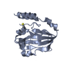
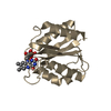
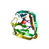
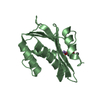
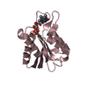
| Title | REFINED STRUCTURES AT 2 ANGSTROMS AND 2.2 ANGSTROMS OF THE TWO FORMS OF THE H-PROTEIN, A LIPOAMIDE-CONTAINING PROTEIN OF THE GLYCINE DECARBOXYLASE COMPLEX | ||||||
 Components Components | H-PROTEIN | ||||||
 Keywords Keywords | OXIDOREDUCTASES(ACTING ON CH-NH2 DONOR) | ||||||
| Function / homology |  Function and homology information Function and homology information glycine cleavage complex / glycine decarboxylation via glycine cleavage system / mitochondrion glycine cleavage complex / glycine decarboxylation via glycine cleavage system / mitochondrionSimilarity search - Function | ||||||
| Biological species |   Pisum sativum (garden pea) Pisum sativum (garden pea) | ||||||
| Method | X-RAY DIFFRACTION / Resolution: 2.2 Å | ||||||
 Authors Authors | Pares, S. / Cohen-Addad, C. | ||||||
 Citation Citation | Journal: Nat.Struct.Biol. / Year: 1995 Title: The lipoamide arm in the glycine decarboxylase complex is not freely swinging. Authors: Cohen-Addad, C. / Pares, S. / Sieker, L. / Neuburger, M. / Douce, R. #1: Journal: Proc.Natl.Acad.Sci.USA / Year: 1994Title: X-Ray Structure Determination at 2.6 Angstroms Resolution of a Lipoate-Containing Protein: The H-Protein of the Glycine Decarboxylase Complex from Pea Leaves Authors: Pares, S. / Cohen-Addad, C. / Sieker, L. / Neuburger, M. / Douce, R. | ||||||
| History |
|
- Structure visualization
| Structure viewer | Molecule: MolmilJmol/JSmol |
|---|
- Downloads & links
-Download
| PDBx/mmCIF format | 1htp.cif.gz | 44.7 KB | Display | PDBx/mmCIF format |
|---|---|---|---|---|
| PDB format | pdb1htp.ent.gz | 30.9 KB | Display | PDB format |
| PDBx/mmJSON format | 1htp.json.gz | Tree view | PDBx/mmJSON format | |
| Others |  Other downloads Other downloads |
-Validation report
| Arichive directory | https://data.pdbj.org/pub/pdb/validation_reports/ht/1htpftp://data.pdbj.org/pub/pdb/validation_reports/ht/1htp | HTTPS FTP |
|---|
-Related structure data
| Similar structure data |
|---|


-Links
 PDBj
PDBj

- Assembly
| Deposited unit | 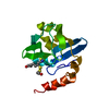
| ||||||||
|---|---|---|---|---|---|---|---|---|---|
| 1 |
| ||||||||
| Unit cell |
|
-Components
| #1: Protein | Mass: 13962.464 Da / Num. of mol.: 1 Source method: isolated from a genetically manipulated source Source: (gene. exp.) Pisum sativum (garden pea)References: UniProt: P16048, glycine dehydrogenase (aminomethyl-transferring) |
|---|---|
| #2: Chemical | ChemComp-OSS /   Mass: 313.500 Da / Num. of mol.: 1 / Source method: obtained synthetically / Formula: C11H23NO3S3 Mass: 313.500 Da / Num. of mol.: 1 / Source method: obtained synthetically / Formula: C11H23NO3S3 |
| #3: Water | ChemComp-HOH / Water Mass: 18.015 Da / Num. of mol.: 145 / Source method: isolated from a natural source / Formula: H2O Mass: 18.015 Da / Num. of mol.: 145 / Source method: isolated from a natural source / Formula: H2O |
| Compound details | COMPND H-PROTEIN CHARGED IN METHYLAMINE OF THE GLYCINE DECARBOXYLASE COMPLEX FROM PEA THE H-PROTEIN ...COMPND H-PROTEIN CHARGED IN METHYLAMIN |
| Sequence details | SEQUENCE REFERENCE DATABASE: SWISS-PROT ENTRY_NAME: GCSH_PEA REFERENCE: MACHEREL D., LEBRUN M. ...SEQUENCE REFERENCE DATABASE: SWISS-PROT ENTRY_NAME: GCSH_PEA REFERENCE: MACHEREL D., LEBRUN M.,GAGNON J., NEUBURGER M., DOUCE R. BIOCHEM J. 268:783-789(1990) KIM Y., OLIVER D.J J. BIOL. CHEM. 265: 848-853(1990). |
-Experimental details
-Experiment
| Experiment | Method: X-RAY DIFFRACTION |
|---|
- Sample preparation
| Crystal | Density Matthews: 2.05 Å3/Da / Density % sol: 40.08 % | ||||||||||||||||||||||||
|---|---|---|---|---|---|---|---|---|---|---|---|---|---|---|---|---|---|---|---|---|---|---|---|---|---|
| Crystal grow | *PLUS Temperature: 8 ℃ / pH: 5.2 / Method: vapor diffusion | ||||||||||||||||||||||||
| Components of the solutions | *PLUS
|
-Data collection
| Radiation | Scattering type: x-ray |
|---|---|
| Radiation wavelength | Relative weight: 1 |
| Reflection | Num. obs: 5684 / % possible obs: 91 % |
| Reflection | *PLUS Highest resolution: 2.2 Å / Num. measured all: 14236 / Rmerge(I) obs: 0.054 |
- Processing
| Software |
| ||||||||||||||||||||||||||||||||||||||||||||||||||||||||||||
|---|---|---|---|---|---|---|---|---|---|---|---|---|---|---|---|---|---|---|---|---|---|---|---|---|---|---|---|---|---|---|---|---|---|---|---|---|---|---|---|---|---|---|---|---|---|---|---|---|---|---|---|---|---|---|---|---|---|---|---|---|---|
| Refinement | Resolution: 2.2→8 Å / σ(F): 2
| ||||||||||||||||||||||||||||||||||||||||||||||||||||||||||||
| Displacement parameters | Biso mean: 17.5 Å2 | ||||||||||||||||||||||||||||||||||||||||||||||||||||||||||||
| Refine analyze | Luzzati coordinate error obs: 0.2 Å | ||||||||||||||||||||||||||||||||||||||||||||||||||||||||||||
| Refinement step | Cycle: LAST / Resolution: 2.2→8 Å
| ||||||||||||||||||||||||||||||||||||||||||||||||||||||||||||
| Refine LS restraints |
|
