 Movie
Movie Controller


+ Open data

- Basic information
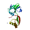
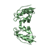
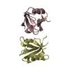
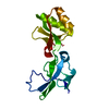
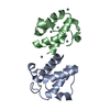
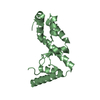
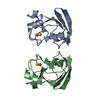
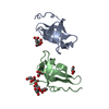
| Entry | Database: PDB / ID: 1ht9 | ||||||
|---|---|---|---|---|---|---|---|
| Title | DOMAIN SWAPPING EF-HANDS | ||||||
 Components Components | CALBINDIN D9K | ||||||
 Keywords Keywords | METAL BINDING PROTEIN / DOMAIN SWAPPING / CALBINDIN D9K /  EF-HAND / CALCIUM BINDING / FOLDING EF-HAND / CALCIUM BINDING / FOLDING | ||||||
| Function / homology |  Function and homology informationvitamin D binding / calcium-dependent protein binding / collagen-containing extracellular matrix / calcium ion binding / extracellular space / cytoplasm Function and homology informationvitamin D binding / calcium-dependent protein binding / collagen-containing extracellular matrix / calcium ion binding / extracellular space / cytoplasmSimilarity search - Function | ||||||
| Biological species |  Bos taurus (cattle) Bos taurus (cattle) | ||||||
| Method | X-RAY DIFFRACTION / SIRAS, MOLECULAR REPLACEMENT / Resolution: 1.76 Å | ||||||
 Authors Authors | Hakansson, M. / Svensson, A.L. / Fast, J. / Linse, S. | ||||||
 Citation Citation | Journal: Protein Sci. / Year: 2001 Title: An extended hydrophobic core induces EF-hand swapping. Authors: Hakansson, M. / Svensson, A. / Fast, J. / Linse, S. #1: Journal: To be PublishedTitle: Symmetric Stabilization of Bound Ca2+ Ions Through Hydrogen Bonding of Coordinating Water Molecules. Ca2+ Binding Abd Structural Stability of Calbindin D9K Mutants Authors: Fast, J. / Muranyi, A. / Gippert, G.P. / Thulin, E. / Evenas, J. / Linse, S. / Hakansson, M. / Svensson, A.L. | ||||||
| History |
|
- Structure visualization
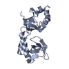
| Structure viewer | Molecule: MolmilJmol/JSmol |
|---|
- Downloads & links
-Download
| PDBx/mmCIF format | 1ht9.cif.gz | 44.9 KB | Display | PDBx/mmCIF format |
|---|---|---|---|---|
| PDB format | pdb1ht9.ent.gz | 31.5 KB | Display | PDB format |
| PDBx/mmJSON format | 1ht9.json.gz | Tree view | PDBx/mmJSON format | |
| Others |  Other downloads Other downloads |
-Validation report
| Arichive directory | https://data.pdbj.org/pub/pdb/validation_reports/ht/1ht9ftp://data.pdbj.org/pub/pdb/validation_reports/ht/1ht9 | HTTPS FTP |
|---|
-Related structure data
| Related structure data |  4icbS S: Starting model for refinement |
|---|---|
| Similar structure data |
-Links
 PDBj
PDBj- Assembly
| Deposited unit | 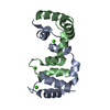
| ||||||||
|---|---|---|---|---|---|---|---|---|---|
| 1 |
| ||||||||
| Unit cell |
|
-Components
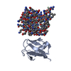
| #1: Protein | Mass: 8661.807 Da / Num. of mol.: 2 / Mutation: YES Source method: isolated from a genetically manipulated source Source: (gene. exp.) Bos taurus (cattle) / Plasmid: PICB1 / Production host:  Escherichia coli (E. coli) / Strain (production host): MM294 / References: UniProt: P02633 Escherichia coli (E. coli) / Strain (production host): MM294 / References: UniProt: P02633#2: Chemical | ChemComp-CA /   Mass: 40.078 Da / Num. of mol.: 4 / Source method: obtained synthetically / Formula: Ca Mass: 40.078 Da / Num. of mol.: 4 / Source method: obtained synthetically / Formula: Ca#3: Water | ChemComp-HOH / | Water Mass: 18.015 Da / Num. of mol.: 86 / Source method: isolated from a natural source / Formula: H2O Mass: 18.015 Da / Num. of mol.: 86 / Source method: isolated from a natural source / Formula: H2O |
|---|
-Experimental details
-Experiment
| Experiment | Method: X-RAY DIFFRACTION / Number of used crystals: 1 |
|---|
- Sample preparation
| Crystal | Density Matthews: 1.87 Å3/Da / Density % sol: 34.25 % | ||||||||||||||||||||||||
|---|---|---|---|---|---|---|---|---|---|---|---|---|---|---|---|---|---|---|---|---|---|---|---|---|---|
| Crystal grow | Temperature: 293 K / Method: evaporation Details: ammonium sulphate, calcium chloride, magnesium chloride, EVAPORATION, temperature 293K | ||||||||||||||||||||||||
| Crystal grow | *PLUS pH: 5.5 / Method: vapor diffusion, hanging drop | ||||||||||||||||||||||||
| Components of the solutions | *PLUS
|
-Data collection
| Diffraction | Mean temperature: 293 K |
|---|---|
| Diffraction source | Source: ROTATING ANODE / Type: RIGAKU RU200 / Wavelength: 1.5418 |
| Detector | Type: MARRESEARCH / Detector: IMAGE PLATE / Date: Sep 9, 1997 |
| Radiation | Monochromator: GRAPHITE / Protocol: SINGLE WAVELENGTH / Monochromatic (M) / Laue (L): M / Scattering type: x-ray |
| Radiation wavelength | Wavelength: 1.5418 Å / Relative weight: 1 |
| Reflection | Resolution: 1.75→44 Å / Num. obs: 50352 / % possible obs: 91.5 % / Observed criterion σ(I): 0 / Redundancy: 4.2 % / Biso Wilson estimate: 18 Å2 / Rmerge(I) obs: 0.06 / Net I/σ(I): 14.5 |
| Reflection shell | Resolution: 1.75→1.8 Å / Redundancy: 3.6 % / Rmerge(I) obs: 0.311 / Mean I/σ(I) obs: 3.7 / % possible all: 60 |
| Reflection | *PLUS Lowest resolution: 44 Å / Num. obs: 11923 / Num. measured all: 50352 |
| Reflection shell | *PLUS % possible obs: 60.1 % |
- Processing
| Software |
| ||||||||||||||||||||
|---|---|---|---|---|---|---|---|---|---|---|---|---|---|---|---|---|---|---|---|---|---|
| Refinement | Method to determine structure: SIRAS, MOLECULAR REPLACEMENT Starting model: PDB ENTRY 4ICB Resolution: 1.76→20 Å / Cross valid method: THROUGHOUT / σ(F): 0
| ||||||||||||||||||||
| Displacement parameters | Biso mean: 23.9 Å2 | ||||||||||||||||||||
| Refinement step | Cycle: LAST / Resolution: 1.76→20 Å
| ||||||||||||||||||||
| Refine LS restraints |
| ||||||||||||||||||||
| Software | *PLUS Name: REFMAC / Classification: refinement | ||||||||||||||||||||
| Refinement | *PLUS % reflection Rfree: 5 % / Rfactor obs: 0.162 | ||||||||||||||||||||
| Solvent computation | *PLUS | ||||||||||||||||||||
| Displacement parameters | *PLUS | ||||||||||||||||||||
| Refine LS restraints | *PLUS
|
