 Movie
Movie Controller

[English] 日本語
 Yorodumi
- PDB-1hm5: CRYSTAL STRUCTURE ANALYSIS OF THE RABBIT D-GLUCOSE 6-PHOSPHATE IS... -
+ Open data

- Basic information
| Entry | Database: PDB / ID: 1hm5 | ||||||
|---|---|---|---|---|---|---|---|
| Title | CRYSTAL STRUCTURE ANALYSIS OF THE RABBIT D-GLUCOSE 6-PHOSPHATE ISOMERASE (NO LIGAND BOUND) | ||||||
 Components Components | PHOSPHOGLUCOSE ISOMERASE Glucose-6-phosphate isomerase Glucose-6-phosphate isomerase | ||||||
 Keywords Keywords | ISOMERASE / dimer | ||||||
| Function / homology |  Function and homology informationglucose-6-phosphate isomerase / glucose-6-phosphate isomerase activity / glucose 6-phosphate metabolic process / carbohydrate derivative binding / monosaccharide binding / cytokine activity / gluconeogenesis / glycolytic process / extracellular space / cytosol Function and homology informationglucose-6-phosphate isomerase / glucose-6-phosphate isomerase activity / glucose 6-phosphate metabolic process / carbohydrate derivative binding / monosaccharide binding / cytokine activity / gluconeogenesis / glycolytic process / extracellular space / cytosolSimilarity search - Function | ||||||
| Biological species |  Oryctolagus cuniculus (rabbit) Oryctolagus cuniculus (rabbit) | ||||||
| Method | X-RAY DIFFRACTION / SYNCHROTRON / MOLECULAR REPLACEMENT / Resolution: 1.8 Å | ||||||
 Authors Authors | Arsenieva, D.A. / Jeffery, C.J. / Hardre, R. / Salmon, L. | ||||||
 Citation Citation | Journal: J.Mol.Biol. / Year: 2002 Title: Conformational Changes in Phosphoglucose Isomerase Induced by Ligand Binding Authors: Arsenieva, D.A. / Jeffery, C.J. | ||||||
| History |
|
- Structure visualization
| Structure viewer | Molecule: MolmilJmol/JSmol |
|---|
- Downloads & links
-Download
| PDBx/mmCIF format | 1hm5.cif.gz | 246 KB | Display | PDBx/mmCIF format |
|---|---|---|---|---|
| PDB format | pdb1hm5.ent.gz | 196.2 KB | Display | PDB format |
| PDBx/mmJSON format | 1hm5.json.gz | Tree view | PDBx/mmJSON format | |
| Others |  Other downloads Other downloads |
-Validation report
| Arichive directory | https://data.pdbj.org/pub/pdb/validation_reports/hm/1hm5ftp://data.pdbj.org/pub/pdb/validation_reports/hm/1hm5 | HTTPS FTP |
|---|
-Related structure data
| Related structure data |  1dqrS S: Starting model for refinement |
|---|---|
| Similar structure data |


-Links
 PDBj
PDBj

- Assembly
| Deposited unit | 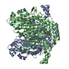
| ||||||||||
|---|---|---|---|---|---|---|---|---|---|---|---|
| 1 |
| ||||||||||
| Unit cell |
| ||||||||||
| Components on special symmetry positions |
| ||||||||||
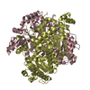
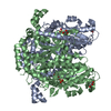
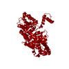
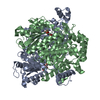
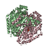
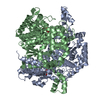
| Details | The active enzyme is a dimer. A complete dimer is in the asymmetric unit. |
-Components
| #1: Protein | Glucose-6-phosphate isomerase / E.C.5.3.1.9 / D-GLUCOSE 6-PHOSPHATE ISOMERASE Mass: 62827.621 Da / Num. of mol.: 2 / Source method: isolated from a natural source / Details: RABBIT SKELETAL MUSCLE TISSUE / Source: (natural) Oryctolagus cuniculus (rabbit) / References: UniProt: Q9N1E2, glucose-6-phosphate isomerase#2: Water | ChemComp-HOH / | Water Mass: 18.015 Da / Num. of mol.: 926 / Source method: isolated from a natural source / Formula: H2O Mass: 18.015 Da / Num. of mol.: 926 / Source method: isolated from a natural source / Formula: H2O |
|---|
-Experimental details
-Experiment
| Experiment | Method: X-RAY DIFFRACTION / Number of used crystals: 1 |
|---|
- Sample preparation
| Crystal | Density Matthews: 2.59 Å3/Da / Density % sol: 52.59 % | |||||||||||||||||||||||||||||||||||||||||||||||||
|---|---|---|---|---|---|---|---|---|---|---|---|---|---|---|---|---|---|---|---|---|---|---|---|---|---|---|---|---|---|---|---|---|---|---|---|---|---|---|---|---|---|---|---|---|---|---|---|---|---|---|
| Crystal grow | Temperature: 295 K / Method: vapor diffusion, hanging drop / pH: 6.6 Details: 14% PEG 8000, 0.1M sodium cacodylate, 0.25M magnesium chloride, pH 6.6 mixed 1:1 with protein in solution 10mM imidazole, 50mM potassium chloride, pH7.5, VAPOR DIFFUSION, HANGING DROP at 295K | |||||||||||||||||||||||||||||||||||||||||||||||||
| Crystal grow | *PLUS pH: 7.5 | |||||||||||||||||||||||||||||||||||||||||||||||||
| Components of the solutions | *PLUS
|
-Data collection
| Diffraction | Mean temperature: 100 K |
|---|---|
| Diffraction source | Source: SYNCHROTRON / Site: APS  / Beamline: 14-BM-C / Wavelength: 1 Å / Beamline: 14-BM-C / Wavelength: 1 Å |
| Detector | Type: ADSC QUANTUM 4 / Detector: CCD / Date: Nov 22, 2000 / Details: mirrors |
| Radiation | Monochromator: bent conical Si-mirror (Rh coating) bend cylindrical Ge(111) monochromator Protocol: SINGLE WAVELENGTH / Monochromatic (M) / Laue (L): M / Scattering type: x-ray |
| Radiation wavelength | Wavelength: 1 Å / Relative weight: 1 |
| Reflection | Resolution: 1.8→40 Å / Num. all: 105425 / Num. obs: 105425 / % possible obs: 87.2 % / Observed criterion σ(F): 0 / Observed criterion σ(I): 0 / Redundancy: 3.8 % / Biso Wilson estimate: 25 Å2 / Rmerge(I) obs: 0.033 / Net I/σ(I): 25.4 |
| Reflection shell | Resolution: 1.8→1.86 Å / Redundancy: 1.8 % / Rmerge(I) obs: 0.258 / Mean I/σ(I) obs: 3.72 / Num. unique all: 5684 / % possible all: 47.5 |
| Reflection | *PLUS Lowest resolution: 40 Å / Num. measured all: 398627 / Rmerge(I) obs: 0.033 |
| Reflection shell | *PLUS % possible obs: 47.5 % / Rmerge(I) obs: 0.258 / Mean I/σ(I) obs: 3.7 |
- Processing
| Software |
| |||||||||||||||||||||||||
|---|---|---|---|---|---|---|---|---|---|---|---|---|---|---|---|---|---|---|---|---|---|---|---|---|---|---|
| Refinement | Method to determine structure: MOLECULAR REPLACEMENT Starting model: PDB ENTRY 1DQR Resolution: 1.8→10 Å / Isotropic thermal model: none / Cross valid method: THROUGHOUT / σ(F): 0 / σ(I): 0 / Stereochemistry target values: CNS
| |||||||||||||||||||||||||
| Refine analyze |
| |||||||||||||||||||||||||
| Refinement step | Cycle: LAST / Resolution: 1.8→10 Å
| |||||||||||||||||||||||||
| Refine LS restraints |
| |||||||||||||||||||||||||
| LS refinement shell | Resolution: 1.8→1.86 Å
| |||||||||||||||||||||||||
| Refinement | *PLUS Lowest resolution: 10 Å / % reflection Rfree: 10 % / Rfactor all: 0.186 / Rfactor obs: 0.183 / Rfactor Rfree: 0.212 / Rfactor Rwork: 0.183 | |||||||||||||||||||||||||
| Solvent computation | *PLUS | |||||||||||||||||||||||||
| Displacement parameters | *PLUS | |||||||||||||||||||||||||
| Refine LS restraints | *PLUS
| |||||||||||||||||||||||||
| LS refinement shell | *PLUS Rfactor Rfree: 0.277 / Rfactor Rwork: 0.252 |
