 Movie
Movie Controller

[English] 日本語
 Yorodumi
- PDB-1hef: The crystal structures at 2.2 angstroms resolution of hydroxyethy... -
+ Open data

- Basic information
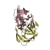
| Entry | Database: PDB / ID: 1hef | |||||||||
|---|---|---|---|---|---|---|---|---|---|---|
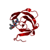
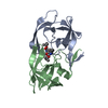
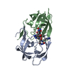
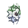
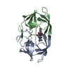
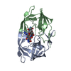
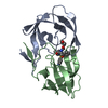
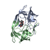
| Title | The crystal structures at 2.2 angstroms resolution of hydroxyethylene-based inhibitors bound to human immunodeficiency virus type 1 protease show that the inhibitors are present in two distinct orientations | |||||||||
 Components Components |
| |||||||||
 Keywords Keywords | HYDROLASE/HYDROLASE INHIBITOR / HYDROLASE-HYDROLASE INHIBITOR COMPLEX | |||||||||
| Function / homology |  Function and homology information Function and homology information HIV-1 retropepsin / : / retroviral ribonuclease H / exoribonuclease H / : / exoribonuclease H activity / host multivesicular body / DNA integration / RNA-directed DNA polymerase / viral genome integration into host DNA ...HIV-1 retropepsin / : / retroviral ribonuclease H / exoribonuclease H / : / exoribonuclease H activity / host multivesicular body / DNA integration / RNA-directed DNA polymerase / viral genome integration into host DNA / viral penetration into host nucleus / establishment of integrated proviral latency / RNA-directed DNA polymerase activity / Transferases; Transferring phosphorus-containing groups; Nucleotidyltransferases / RNA-DNA hybrid ribonuclease activity / viral nucleocapsid / DNA recombination / Hydrolases; Acting on ester bonds / DNA-directed DNA polymerase / aspartic-type endopeptidase activity / DNA-directed DNA polymerase activity / symbiont entry into host cell / symbiont-mediated suppression of host gene expression / lipid binding / host cell nucleus / host cell plasma membrane / virion membrane / structural molecule activity / proteolysis / DNA binding / RNA binding / zinc ion binding / membrane HIV-1 retropepsin / : / retroviral ribonuclease H / exoribonuclease H / : / exoribonuclease H activity / host multivesicular body / DNA integration / RNA-directed DNA polymerase / viral genome integration into host DNA ...HIV-1 retropepsin / : / retroviral ribonuclease H / exoribonuclease H / : / exoribonuclease H activity / host multivesicular body / DNA integration / RNA-directed DNA polymerase / viral genome integration into host DNA / viral penetration into host nucleus / establishment of integrated proviral latency / RNA-directed DNA polymerase activity / Transferases; Transferring phosphorus-containing groups; Nucleotidyltransferases / RNA-DNA hybrid ribonuclease activity / viral nucleocapsid / DNA recombination / Hydrolases; Acting on ester bonds / DNA-directed DNA polymerase / aspartic-type endopeptidase activity / DNA-directed DNA polymerase activity / symbiont entry into host cell / symbiont-mediated suppression of host gene expression / lipid binding / host cell nucleus / host cell plasma membrane / virion membrane / structural molecule activity / proteolysis / DNA binding / RNA binding / zinc ion binding / membraneSimilarity search - Function | |||||||||
| Biological species |   Human immunodeficiency virus 1 Human immunodeficiency virus 1 | |||||||||
| Method | X-RAY DIFFRACTION / Resolution: 2.2 Å | |||||||||
 Authors Authors | Murthy, K. / Winborne, E.L. / Minnich, M.D. / Culp, J.S. / Debouck, C. | |||||||||
 Citation Citation | Journal: J.Biol.Chem. / Year: 1992 Title: The crystal structures at 2.2-A resolution of hydroxyethylene-based inhibitors bound to human immunodeficiency virus type 1 protease show that the inhibitors are present in two distinct orientations. Authors: Murthy, K.H. / Winborne, E.L. / Minnich, M.D. / Culp, J.S. / Debouck, C. | |||||||||
| History |
| |||||||||
| Remark 300 | HIV-1 PROTEASE IS A SYMMETRIC DIMER, WHILE THE INHIBITOR IS AN ASYMMETRIC MOLECULE. FURTHERMORE, ...HIV-1 PROTEASE IS A SYMMETRIC DIMER, WHILE THE INHIBITOR IS AN ASYMMETRIC MOLECULE. FURTHERMORE, THE INHIBITOR IS SMALL ENOUGH TO BE ENTIRELY CONTAINED WITHIN THE ACTIVE SITE OF THE ENZYME AND, THEREFORE, DOES NOT CONTRIBUTE TO INTER-DIMER CONTACTS. THUS, OF THE TWO POSSIBLE ORIENTATIONS OF THE INHIBITOR, NEITHER IS THERMODYNAMICALLY PREFERRED. IN THE CRYSTAL STRUCTURE, THEREFORE, BOTH ARE REPRESENTED EQUALLY. THE ASYMMETRIC UNIT OF THE CRYSTAL THUS CONSISTS OF ONE PROTEASE MONOMER, AND ONE COPY OF EACH OF TWO POSSIBLE ORIENTATIONS OF THE INHIBITOR. EACH COPY OF THE INHIBITOR REPRESENTS HALF THE TOTAL OCCUPANCY FOR THE INHIBITOR. |
- Structure visualization
| Structure viewer | Molecule: MolmilJmol/JSmol |
|---|
- Downloads & links
-Download
| PDBx/mmCIF format | 1hef.cif.gz | 36 KB | Display | PDBx/mmCIF format |
|---|---|---|---|---|
| PDB format | pdb1hef.ent.gz | 22.8 KB | Display | PDB format |
| PDBx/mmJSON format | 1hef.json.gz | Tree view | PDBx/mmJSON format | |
| Others |  Other downloads Other downloads |
-Validation report
| Arichive directory | https://data.pdbj.org/pub/pdb/validation_reports/he/1hefftp://data.pdbj.org/pub/pdb/validation_reports/he/1hef | HTTPS FTP |
|---|
-Related structure data


-Links
 PDBj
PDBj


- Assembly
| Deposited unit | 
| ||||||||
|---|---|---|---|---|---|---|---|---|---|
| 1 | 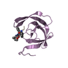
| ||||||||
| Unit cell |
| ||||||||
| Components on special symmetry positions |
|
-Components
| #1: Protein | Mass: 10786.663 Da / Num. of mol.: 1 Source method: isolated from a genetically manipulated source Source: (gene. exp.) Human immunodeficiency virus 1 / Genus: Lentivirus / Production host:  Escherichia coli (E. coli) / References: UniProt: P03366 Escherichia coli (E. coli) / References: UniProt: P03366 |
|---|---|
| #2: Protein/peptide |   Type: Peptide-like / Class: Inhibitor / Mass: 667.835 Da / Num. of mol.: 1 / Source method: obtained synthetically Type: Peptide-like / Class: Inhibitor / Mass: 667.835 Da / Num. of mol.: 1 / Source method: obtained syntheticallyReferences: methyl N-{(2S,4S,5S)-5-[(L-alanyl-L-alanyl)amino]-2-benzyl-4-hydroxy-6-phenylhexanoyl}-L-valyl-L-valinate |
| #3: Water | ChemComp-HOH / Water Mass: 18.015 Da / Num. of mol.: 23 / Source method: isolated from a natural source / Formula: H2O Mass: 18.015 Da / Num. of mol.: 23 / Source method: isolated from a natural source / Formula: H2O |
| Sequence details | THIS VARIANT OF THE HIV 1 PROTEASE HAS ASN AT THE POSITION 36 IN CHAIN E AS CONFIRMED BY INSPECTION ...THIS VARIANT OF THE HIV 1 PROTEASE HAS ASN AT THE POSITION 36 IN CHAIN E AS CONFIRMED BY INSPECTION |
-Experimental details
-Experiment
| Experiment | Method: X-RAY DIFFRACTION |
|---|
- Sample preparation
| Crystal | Density Matthews: 2.09 Å3/Da / Density % sol: 41.08 % | ||||||||||||||||||||||||||||||||||||||||||||||||
|---|---|---|---|---|---|---|---|---|---|---|---|---|---|---|---|---|---|---|---|---|---|---|---|---|---|---|---|---|---|---|---|---|---|---|---|---|---|---|---|---|---|---|---|---|---|---|---|---|---|
| Crystal grow | *PLUS pH: 5 / Method: vapor diffusion, hanging drop | ||||||||||||||||||||||||||||||||||||||||||||||||
| Components of the solutions | *PLUS
|
-Data collection
| Radiation | Scattering type: x-ray |
|---|---|
| Radiation wavelength | Relative weight: 1 |
- Processing
| Software | Name: PROLSQ / Classification: refinement | ||||||||||||||||||||||||||||||||||||||||||||||||||||||||||||||||||||||||||||||||||||
|---|---|---|---|---|---|---|---|---|---|---|---|---|---|---|---|---|---|---|---|---|---|---|---|---|---|---|---|---|---|---|---|---|---|---|---|---|---|---|---|---|---|---|---|---|---|---|---|---|---|---|---|---|---|---|---|---|---|---|---|---|---|---|---|---|---|---|---|---|---|---|---|---|---|---|---|---|---|---|---|---|---|---|---|---|---|
| Refinement | Resolution: 2.2→5 Å / σ(I): 2 Details: THE INHIBITOR ALA-ALA-PJJ-VAL-VME IS DISORDERED AROUND THE TWO-FOLD CYRSTALLOGRAPHIC AXIS. APPLICATION OF CRYSTALLOGRAPHIC SYMMETRY RESULTS IN THE OVERLAP OF THE TWO ORIENTATIONS. THE ...Details: THE INHIBITOR ALA-ALA-PJJ-VAL-VME IS DISORDERED AROUND THE TWO-FOLD CYRSTALLOGRAPHIC AXIS. APPLICATION OF CRYSTALLOGRAPHIC SYMMETRY RESULTS IN THE OVERLAP OF THE TWO ORIENTATIONS. THE OCCUPANCY OF EACH ORIENTATION IS 1/2.
| ||||||||||||||||||||||||||||||||||||||||||||||||||||||||||||||||||||||||||||||||||||
| Refinement step | Cycle: LAST / Resolution: 2.2→5 Å
| ||||||||||||||||||||||||||||||||||||||||||||||||||||||||||||||||||||||||||||||||||||
| Refine LS restraints |
| ||||||||||||||||||||||||||||||||||||||||||||||||||||||||||||||||||||||||||||||||||||
| Software | *PLUS Name: PROLSQ / Classification: refinement | ||||||||||||||||||||||||||||||||||||||||||||||||||||||||||||||||||||||||||||||||||||
| Refinement | *PLUS Rfactor obs: 0.159 | ||||||||||||||||||||||||||||||||||||||||||||||||||||||||||||||||||||||||||||||||||||
| Solvent computation | *PLUS | ||||||||||||||||||||||||||||||||||||||||||||||||||||||||||||||||||||||||||||||||||||
| Displacement parameters | *PLUS |
