 Movie
Movie Controller

[English] 日本語
 Yorodumi
- PDB-1hak: CRYSTAL STRUCTURE OF RECOMBINANT HUMAN PLACENTAL ANNEXIN V COMPLE... -
+ Open data

- Basic information
| Entry | Database: PDB / ID: 1hak | ||||||
|---|---|---|---|---|---|---|---|
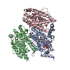
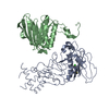
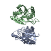
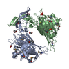
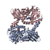
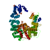
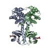
| Title | CRYSTAL STRUCTURE OF RECOMBINANT HUMAN PLACENTAL ANNEXIN V COMPLEXED WITH K-201 AS A CALCIUM CHANNEL ACTIVITY INHIBITOR | ||||||
 Components Components | ANNEXIN V Annexin A5 Annexin A5 | ||||||
 Keywords Keywords | CALCIUM/PHOSPHOLIPID-BINDING / ANNEXIN V / LIPOCORTIN V / ENDONEXIN II / PLACENTA ANTICOAGULANT PROTEIN-I / 35KDA CALELECTRIN / INHIBITOR OF BLOOD COAGULATION / CALCIUM-PHOSPHOLIPID-BINDING complex | ||||||
| Function / homology |  Function and homology information Function and homology informationphospholipase inhibitor activity / endothelial microparticle / negative regulation of coagulation / calcium-dependent phospholipid binding / phosphatidylserine binding / : / phospholipid binding / sarcolemma / blood coagulation / Platelet degranulation ...phospholipase inhibitor activity / endothelial microparticle / negative regulation of coagulation / calcium-dependent phospholipid binding / phosphatidylserine binding / : / phospholipid binding / sarcolemma / blood coagulation / Platelet degranulation / collagen-containing extracellular matrix / external side of plasma membrane / focal adhesion / calcium ion binding / negative regulation of apoptotic process / signal transduction / extracellular exosome / extracellular region / membrane / cytosol / cytoplasmSimilarity search - Function | ||||||
| Biological species |  Homo sapiens (human) Homo sapiens (human) | ||||||
| Method | X-RAY DIFFRACTION / MOLECULAR REPLACEMENT / Resolution: 3 Å | ||||||
 Authors Authors | Ago, H. / Inagaki, E. / Miyano, M. | ||||||
 Citation Citation | Journal: J.Mol.Biol. / Year: 1997 Title: Crystal structure of annexin V with its ligand K-201 as a calcium channel activity inhibitor. Authors: Kaneko, N. / Ago, H. / Matsuda, R. / Inagaki, E. / Miyano, M. #1: Journal: Drug Dev.Res. / Year: 1994Title: New 1,4-Bezothiazepin Derivative, K201, Demonstrates Cardioprotective Effects Against Sudden Cardiac Cell Death and Intracellular Calcium Blocking Action Authors: Kaneko, N. #2: Journal: J.Mol.Biol. / Year: 1992Title: Crystal and Molecular Structure of Human Annexin V After Refinement. Implications for Structure, Membrane Binding and Ion Channel Formation of the Annexin Family of Proteins Authors: Huber, R. / Berendes, R. / Burger, A. / Schneider, M. / Karshikov, A. / Luecke, H. / Romisch, J. / Paques, E. #3: Journal: J.Biochem.(Tokyo) / Year: 1987Title: Structure and Expression of Cdna for an Inhibitor of Blood Coagulation Isolated from Human Placenta: A New Lipocortin-Like Protein Authors: Iwasaki, A. / Suda, M. / Nakao, H. / Nagoya, T. / Saino, Y. / Arai, K. / Mizoguchi, T. / Sato, F. / Yoshizaki, H. / Hirata, M. / Miyata, T. / Shidara, Y. / Murata, M. / Maki, M. | ||||||
| History |
|
- Structure visualization
| Structure viewer | Molecule: MolmilJmol/JSmol |
|---|
- Downloads & links
-Download
| PDBx/mmCIF format | 1hak.cif.gz | 135.4 KB | Display | PDBx/mmCIF format |
|---|---|---|---|---|
| PDB format | pdb1hak.ent.gz | 107.8 KB | Display | PDB format |
| PDBx/mmJSON format | 1hak.json.gz | Tree view | PDBx/mmJSON format | |
| Others |  Other downloads Other downloads |
-Validation report
| Arichive directory | https://data.pdbj.org/pub/pdb/validation_reports/ha/1hakftp://data.pdbj.org/pub/pdb/validation_reports/ha/1hak | HTTPS FTP |
|---|
-Related structure data
| Related structure data |  1ak0S S: Starting model for refinement |
|---|---|
| Similar structure data |

-Links
 PDBj
PDBj- Assembly
| Deposited unit | 
| ||||||||
|---|---|---|---|---|---|---|---|---|---|
| 1 |
| ||||||||
| Unit cell |
|
-Components
| #1: Protein | Annexin A5 / LIPOCORTIN V / ENDONEXIN II / PLACENTA ANTICOAGULANT PROTEIN-I / 35KDA CALELECTRIN / INHIBITOR OF ...LIPOCORTIN V / ENDONEXIN II / PLACENTA ANTICOAGULANT PROTEIN-I / 35KDA CALELECTRIN / INHIBITOR OF BLOOD COAGULATION Mass: 35980.707 Da / Num. of mol.: 2 / Fragment: ANNEXIN FOLD Source method: isolated from a genetically manipulated source Source: (gene. exp.) Homo sapiens (human) / Cell line: 293 / Cellular location: CYTOPLASM / Gene: CDNA / Organ: PLACENTA / Production host:  Escherichia coli (E. coli) / Strain (production host): JM105 / References: UniProt: P08758 Escherichia coli (E. coli) / Strain (production host): JM105 / References: UniProt: P08758#2: Chemical |   Mass: 424.599 Da / Num. of mol.: 2 / Source method: obtained synthetically / Formula: C25H32N2O2S Mass: 424.599 Da / Num. of mol.: 2 / Source method: obtained synthetically / Formula: C25H32N2O2S#3: Water | ChemComp-HOH / | Water Mass: 18.015 Da / Num. of mol.: 109 / Source method: isolated from a natural source / Formula: H2O Mass: 18.015 Da / Num. of mol.: 109 / Source method: isolated from a natural source / Formula: H2O |
|---|
-Experimental details
-Experiment
| Experiment | Method: X-RAY DIFFRACTION / Number of used crystals: 1 |
|---|
- Sample preparation
| Crystal | Density Matthews: 2.6 Å3/Da / Density % sol: 52 % | |||||||||||||||
|---|---|---|---|---|---|---|---|---|---|---|---|---|---|---|---|---|
| Crystal grow | pH: 7.5 / Details: pH 7.5 | |||||||||||||||
| Crystal grow | *PLUS Method: unknown | |||||||||||||||
| Components of the solutions | *PLUS
|
-Data collection
| Diffraction | Mean temperature: 298 K |
|---|---|
| Diffraction source | Source: ROTATING ANODE / Type: RIGAKU RUH3R / Wavelength: 1.5418 |
| Detector | Type: RIGAKU RAXIS IIC / Detector: IMAGE PLATE / Date: Sep 14, 1995 / Details: ASYMMETRIC DOUBLE MIRROR |
| Radiation | Monochromator: NI FILTER / Monochromatic (M) / Laue (L): M / Scattering type: x-ray |
| Radiation wavelength | Wavelength: 1.5418 Å / Relative weight: 1 |
| Reflection | Resolution: 3→40 Å / Num. obs: 104199 / % possible obs: 98.3 % / Observed criterion σ(I): 1 / Redundancy: 4.2 % / Rmerge(I) obs: 0.068 / Net I/σ(I): 17.6 |
- Processing
| Software |
| ||||||||||||||||||||||||||||||||||||||||||||||||||||||||||||
|---|---|---|---|---|---|---|---|---|---|---|---|---|---|---|---|---|---|---|---|---|---|---|---|---|---|---|---|---|---|---|---|---|---|---|---|---|---|---|---|---|---|---|---|---|---|---|---|---|---|---|---|---|---|---|---|---|---|---|---|---|---|
| Refinement | Method to determine structure: MOLECULAR REPLACEMENT Starting model: 1AK0 Resolution: 3→20 Å / Rfactor Rfree error: 0.0125 / Data cutoff high absF: 100000 / Data cutoff low absF: 0.1 / Cross valid method: THROUGHOUT / σ(F): 2 Details: THE PSF FILE FOR X-PLOR REFINEMENT WAS CREATED USING QUANTA/CHARMM.
| ||||||||||||||||||||||||||||||||||||||||||||||||||||||||||||
| Displacement parameters | Biso mean: 12.6 Å2 | ||||||||||||||||||||||||||||||||||||||||||||||||||||||||||||
| Refinement step | Cycle: LAST / Resolution: 3→20 Å
| ||||||||||||||||||||||||||||||||||||||||||||||||||||||||||||
| Refine LS restraints |
| ||||||||||||||||||||||||||||||||||||||||||||||||||||||||||||
| LS refinement shell | Resolution: 3→3.14 Å / Rfactor Rfree error: 0.006 / Total num. of bins used: 8
| ||||||||||||||||||||||||||||||||||||||||||||||||||||||||||||
| Xplor file |
| ||||||||||||||||||||||||||||||||||||||||||||||||||||||||||||
| Software | *PLUS Name: X-PLOR / Version: 3.1F / Classification: refinement | ||||||||||||||||||||||||||||||||||||||||||||||||||||||||||||
| Refinement | *PLUS | ||||||||||||||||||||||||||||||||||||||||||||||||||||||||||||
| Solvent computation | *PLUS | ||||||||||||||||||||||||||||||||||||||||||||||||||||||||||||
| Displacement parameters | *PLUS | ||||||||||||||||||||||||||||||||||||||||||||||||||||||||||||
| Refine LS restraints | *PLUS
| ||||||||||||||||||||||||||||||||||||||||||||||||||||||||||||
| LS refinement shell | *PLUS Rfactor obs: 0.273 |
