 Movie
Movie Controller

[English] 日本語
 Yorodumi
- PDB-1h05: 3-DEHYDROQUINATE DEHYDRATASE FROM MYCOBACTERIUM TUBERCULOSIS IN C... -
+ Open data

- Basic information
| Entry | Database: PDB / ID: 1h05 | ||||||
|---|---|---|---|---|---|---|---|
| Title | 3-DEHYDROQUINATE DEHYDRATASE FROM MYCOBACTERIUM TUBERCULOSIS IN COMPLEX WITH SULPHATE | ||||||
 Components Components | 3-DEHYDROQUINATE DEHYDRATASE | ||||||
 Keywords Keywords | DEHYDRATASE / SHIKIMATE PATHWAY / ALPHA/BETA PROTEIN / LYASE / AROMATIC AMINO ACID BIOSYNTHESIS | ||||||
| Function / homology |  Function and homology information Function and homology informationquinate catabolic process / Chorismate via Shikimate Pathway / 3-dehydroquinate dehydratase / 3-dehydroquinate dehydratase activity / chorismate biosynthetic process / aromatic amino acid family biosynthetic process / amino acid biosynthetic process / cytosolSimilarity search - Function | ||||||
| Biological species |   MYCOBACTERIUM TUBERCULOSIS (bacteria) MYCOBACTERIUM TUBERCULOSIS (bacteria) | ||||||
| Method | X-RAY DIFFRACTION / SYNCHROTRON / MOLECULAR REPLACEMENT / Resolution: 1.5 Å | ||||||
 Authors Authors | Roszak, A.W. / Coggins, J.R. / Lapthorn, A.J. | ||||||
 Citation Citation | Journal: FEBS Lett. / Year: 2002 Title: Specificity of Substrate Recognition by Type II Dehydroquinases as Revealed by Binding of Polyanions(1) Authors: Evans, L. / Roszak, A.W. / Noble, L. / Robinson, D. / Chalk, P. / Matthews, J. / Coggins, J.R. / Price, N. / Lapthorn, A.J. #1: Journal: Nat.Struct.Biol. / Year: 1999Title: The Two Types of 3-Dehydroquinase Have Different Structures But Catalyze the Same Overall Reaction Authors: Gourley, D.G. / Shrive, A.K. / Polikarpov, I. / Krell, T. / Coggins, J.R. / Hawkins, A.R. / Isaacs, N.W. / Sawyer, L. | ||||||
| History |
|
- Structure visualization
| Structure viewer | Molecule: MolmilJmol/JSmol |
|---|
- Downloads & links
-Download
| PDBx/mmCIF format | 1h05.cif.gz | 76.7 KB | Display | PDBx/mmCIF format |
|---|---|---|---|---|
| PDB format | pdb1h05.ent.gz | 58.3 KB | Display | PDB format |
| PDBx/mmJSON format | 1h05.json.gz | Tree view | PDBx/mmJSON format | |
| Others |  Other downloads Other downloads |
-Validation report
| Arichive directory | https://data.pdbj.org/pub/pdb/validation_reports/h0/1h05ftp://data.pdbj.org/pub/pdb/validation_reports/h0/1h05 | HTTPS FTP |
|---|
-Related structure data
| Related structure data |  2dhqS S: Starting model for refinement |
|---|---|
| Similar structure data |


-Links
 PDBj
PDBj
- Assembly
| Deposited unit | 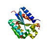
| |||||||||||||||||||||||||||
|---|---|---|---|---|---|---|---|---|---|---|---|---|---|---|---|---|---|---|---|---|---|---|---|---|---|---|---|---|
| 1 | x 12
| |||||||||||||||||||||||||||
| Unit cell |
| |||||||||||||||||||||||||||
| Components on special symmetry positions |
|
-Components
| #1: Protein | / 3-DEHYDROQUINASE / TYPE 2 DHQASE Mass: 15676.737 Da / Num. of mol.: 1 Source method: isolated from a genetically manipulated source Source: (gene. exp.) MYCOBACTERIUM TUBERCULOSIS (bacteria) / Strain: H37RV / Plasmid: MPET / Production host: ESCHERICHIA COLI (E. coli) / Strain (production host): BL21(DE3)References: UniProt: P36918, UniProt: P9WPX7*PLUS, 3-dehydroquinate dehydratase | ||||
|---|---|---|---|---|---|
| #2: Chemical | ChemComp-SO4 / Sulfate  Mass: 96.063 Da / Num. of mol.: 4 / Source method: obtained synthetically / Formula: SO4 Mass: 96.063 Da / Num. of mol.: 4 / Source method: obtained synthetically / Formula: SO4#3: Water | ChemComp-HOH / | Water Mass: 18.015 Da / Num. of mol.: 200 / Source method: isolated from a natural source / Formula: H2O Mass: 18.015 Da / Num. of mol.: 200 / Source method: isolated from a natural source / Formula: H2OCompound details | CATALYZES TRANS-DEHYDRATIO | |
-Experimental details
-Experiment
| Experiment | Method: X-RAY DIFFRACTION / Number of used crystals: 1 |
|---|
- Sample preparation
| Crystal | Density Matthews: 2.77 Å3/Da / Density % sol: 55.5 % Description: LOW COMPLETENESS AND REDUNDANCY IN HIGHEST RESOLUTION SHELL AS DATA WERE PROCESSED TO CORNERS OF CCD DETECTOR | ||||||||||||||||||||||||||||||
|---|---|---|---|---|---|---|---|---|---|---|---|---|---|---|---|---|---|---|---|---|---|---|---|---|---|---|---|---|---|---|---|
| Crystal grow | pH: 7.5 / Details: 30% MPD, 0.5M AMMONIUM SULPHATE 0.1M HEPES PH 7.5 | ||||||||||||||||||||||||||||||
| Crystal grow | *PLUS Method: vapor diffusion, sitting drop | ||||||||||||||||||||||||||||||
| Components of the solutions | *PLUS
|
-Data collection
| Diffraction | Mean temperature: 100 K |
|---|---|
| Diffraction source | Source: SYNCHROTRON / Site: SRS  / Beamline: PX9.6 / Wavelength: 0.87 / Beamline: PX9.6 / Wavelength: 0.87 |
| Detector | Type: ADSC CCD / Detector: CCD / Date: Feb 4, 1999 |
| Radiation | Protocol: SINGLE WAVELENGTH / Monochromatic (M) / Laue (L): M / Scattering type: x-ray |
| Radiation wavelength | Wavelength: 0.87 Å / Relative weight: 1 |
| Reflection | Resolution: 1.5→30 Å / Num. obs: 24274 / % possible obs: 89.9 % / Redundancy: 3.5 % / Rmerge(I) obs: 0.039 / Net I/σ(I): 20.3 |
| Reflection shell | Resolution: 1.5→1.53 Å / Rmerge(I) obs: 0.832 / % possible all: 32.7 |
| Reflection | *PLUS Highest resolution: 1.45 Å / Num. measured all: 85115 |
| Reflection shell | *PLUS % possible obs: 32.7 % |
- Processing
| Software |
| ||||||||||||||||||||
|---|---|---|---|---|---|---|---|---|---|---|---|---|---|---|---|---|---|---|---|---|---|
| Refinement | Method to determine structure: MOLECULAR REPLACEMENT Starting model: PDB ENTRY 2DHQ Resolution: 1.5→30 Å / σ(F): 0 / ESU R: 0.08894 / ESU R Free: 0.07971
| ||||||||||||||||||||
| Displacement parameters | Biso mean: 20.1 Å2 | ||||||||||||||||||||
| Refinement step | Cycle: LAST / Resolution: 1.5→30 Å
| ||||||||||||||||||||
| Refine LS restraints | *PLUS
| ||||||||||||||||||||
| LS refinement shell | *PLUS Highest resolution: 1.5 Å / Lowest resolution: 1.57 Å / Rfactor Rwork: 0.1 / Num. reflection Rwork: 1141 / Total num. of bins used: 20 |
