 Movie
Movie Controller


+ Open data

- Basic information
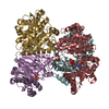
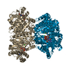
| Entry | Database: PDB / ID: 2dhq | ||||||
|---|---|---|---|---|---|---|---|
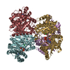
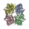
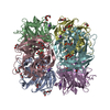
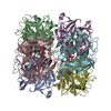
| Title | 3-DEHYDROQUINATE DEHYDRATASE FROM MYCOBACTERIUM TUBERCULOSIS | ||||||
 Components Components | PROTEIN (3-DEHYDROQUINATE DEHYDRATASE) | ||||||
 Keywords Keywords |  DEHYDRATASE / SHIKIMATE PATHWAY / ALPHA/BETA PROTEIN DEHYDRATASE / SHIKIMATE PATHWAY / ALPHA/BETA PROTEIN | ||||||
| Function / homology |  Function and homology information Function and homology informationquinate catabolic process / Chorismate via Shikimate Pathway / 3-dehydroquinate dehydratase / 3-dehydroquinate dehydratase activity / chorismate biosynthetic process / aromatic amino acid family biosynthetic process / amino acid biosynthetic process / cytosolSimilarity search - Function | ||||||
| Biological species |  Mycobacterium tuberculosis H37Rv (bacteria) Mycobacterium tuberculosis H37Rv (bacteria) | ||||||
| Method | X-RAY DIFFRACTION / SYNCHROTRON / MIRAS / Resolution: 2 Å | ||||||
 Authors Authors | Gourley, D.G. / Hawkins, A.R. / Coggins, J.R. / Isaacs, N.W. | ||||||
 Citation Citation | Journal: Nat.Struct.Biol. / Year: 1999 Title: The two types of 3-dehydroquinase have distinct structures but catalyze the same overall reaction. Authors: Gourley, D.G. / Shrive, A.K. / Polikarpov, I. / Krell, T. / Coggins, J.R. / Hawkins, A.R. / Isaacs, N.W. / Sawyer, L. | ||||||
| History |
|
- Structure visualization
| Structure viewer | Molecule: MolmilJmol/JSmol |
|---|
- Downloads & links
-Download
| PDBx/mmCIF format | 2dhq.cif.gz | 41.1 KB | Display | PDBx/mmCIF format |
|---|---|---|---|---|
| PDB format | pdb2dhq.ent.gz | 29.8 KB | Display | PDB format |
| PDBx/mmJSON format | 2dhq.json.gz | Tree view | PDBx/mmJSON format | |
| Others |  Other downloads Other downloads |
-Validation report
| Arichive directory | https://data.pdbj.org/pub/pdb/validation_reports/dh/2dhqftp://data.pdbj.org/pub/pdb/validation_reports/dh/2dhq | HTTPS FTP |
|---|
-Related structure data


-Links
 PDBj
PDBj
- Assembly
| Deposited unit | 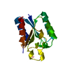
| ||||||||||||
|---|---|---|---|---|---|---|---|---|---|---|---|---|---|
| 1 | x 10
| ||||||||||||
| 2 | x 12
| ||||||||||||
| Unit cell |
| ||||||||||||
| Components on special symmetry positions |
|
-Components
| #1: Protein | Mass: 15676.737 Da / Num. of mol.: 1 Source method: isolated from a genetically manipulated source Source: (gene. exp.) Mycobacterium tuberculosis H37Rv (bacteria)Species: Mycobacterium tuberculosis / Strain: H37RV / Plasmid: PKK44 / Gene (production host): AROQ / Production host: Escherichia coli (E. coli) / Strain (production host): SK3430 / Variant (production host): ARO D-References: UniProt: P0A4Z6, UniProt: P9WPX7*PLUS, 3-dehydroquinate dehydratase |
|---|---|
| #2: Water | ChemComp-HOH / Water Mass: 18.015 Da / Num. of mol.: 123 / Source method: isolated from a natural source / Formula: H2O Mass: 18.015 Da / Num. of mol.: 123 / Source method: isolated from a natural source / Formula: H2O |
-Experimental details
-Experiment
| Experiment | Method: X-RAY DIFFRACTION / Number of used crystals: 2 |
|---|
- Sample preparation
| Crystal | Density Matthews: 2.77 Å3/Da / Density % sol: 55.5 % | |||||||||||||||||||||||||
|---|---|---|---|---|---|---|---|---|---|---|---|---|---|---|---|---|---|---|---|---|---|---|---|---|---|---|
| Crystal grow | pH: 7.2 / Details: pH 7.20 | |||||||||||||||||||||||||
| Crystal | *PLUS Density % sol: 54 % | |||||||||||||||||||||||||
| Crystal grow | *PLUS pH: 7.16 / Method: vapor diffusion, sitting dropDetails: Gourley, D.G., (1994) Nature Struct. Biol., 241, 488. | |||||||||||||||||||||||||
| Components of the solutions | *PLUS
|
-Data collection
| Diffraction | Mean temperature: 287 K |
|---|---|
| Diffraction source | Source: SYNCHROTRON / Site: SRS  / Beamline: PX9.5 / Wavelength: 0.89 / Beamline: PX9.5 / Wavelength: 0.89 |
| Detector | Type: MARRESEARCH / Detector: AREA DETECTOR / Details: MIRRORS |
| Radiation | Protocol: SINGLE WAVELENGTH / Monochromatic (M) / Laue (L): M / Scattering type: x-ray |
| Radiation wavelength | Wavelength: 0.89 Å / Relative weight: 1 |
| Reflection | Resolution: 2→30 Å / Num. obs: 11412 / % possible obs: 97.4 % / Redundancy: 3.3 % / Biso Wilson estimate: 19.55 Å2 / Rmerge(I) obs: 0.062 / Net I/σ(I): 7.44 |
| Reflection shell | Resolution: 2→2.11 Å / Redundancy: 2.7 % / Rmerge(I) obs: 0.102 / Mean I/σ(I) obs: 7 / % possible all: 94.6 |
| Reflection | *PLUS Num. measured all: 37418 |
- Processing
| Software |
| ||||||||||||||||||||||||||||||||||||||||||||||||||||||||||||||||||||||||||||||||||||
|---|---|---|---|---|---|---|---|---|---|---|---|---|---|---|---|---|---|---|---|---|---|---|---|---|---|---|---|---|---|---|---|---|---|---|---|---|---|---|---|---|---|---|---|---|---|---|---|---|---|---|---|---|---|---|---|---|---|---|---|---|---|---|---|---|---|---|---|---|---|---|---|---|---|---|---|---|---|---|---|---|---|---|---|---|---|
| Refinement | Method to determine structure: MIRAS / Resolution: 2→30 Å / Cross valid method: THROUGHOUT / σ(F): 0 Details: THR: THE C-TERMINAL RESIDUE WAS NOT SEEN IN THE DENSITY MAP
| ||||||||||||||||||||||||||||||||||||||||||||||||||||||||||||||||||||||||||||||||||||
| Displacement parameters | Biso mean: 20.1 Å2 | ||||||||||||||||||||||||||||||||||||||||||||||||||||||||||||||||||||||||||||||||||||
| Refinement step | Cycle: LAST / Resolution: 2→30 Å
| ||||||||||||||||||||||||||||||||||||||||||||||||||||||||||||||||||||||||||||||||||||
| Refine LS restraints |
| ||||||||||||||||||||||||||||||||||||||||||||||||||||||||||||||||||||||||||||||||||||
| Software | *PLUS Name: REFMAC / Classification: refinement | ||||||||||||||||||||||||||||||||||||||||||||||||||||||||||||||||||||||||||||||||||||
| Refinement | *PLUS Highest resolution: 2 Å / Lowest resolution: 30 Å / σ(F): 0 / % reflection Rfree: 10 % / Rfactor obs: 0.147 | ||||||||||||||||||||||||||||||||||||||||||||||||||||||||||||||||||||||||||||||||||||
| Solvent computation | *PLUS | ||||||||||||||||||||||||||||||||||||||||||||||||||||||||||||||||||||||||||||||||||||
| Displacement parameters | *PLUS Biso mean: 20.1 Å2 | ||||||||||||||||||||||||||||||||||||||||||||||||||||||||||||||||||||||||||||||||||||
| Refine LS restraints | *PLUS
|
