 Movie
Movie Controller


+ Open data

- Basic information
| Entry | Database: PDB / ID: 1gv4 | ||||||
|---|---|---|---|---|---|---|---|
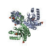
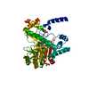
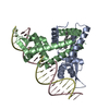
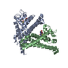
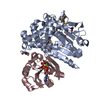
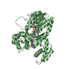
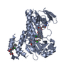
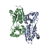
| Title | Murine apoptosis-inducing factor (AIF) | ||||||
 Components Components | PROGRAMED CELL DEATH PROTEIN 8 | ||||||
 Keywords Keywords |  OXIDOREDUCTASE / FLAVOPROTEIN / FAD / NUCLEAR PROTEIN / APOPTOSI OXIDOREDUCTASE / FLAVOPROTEIN / FAD / NUCLEAR PROTEIN / APOPTOSI | ||||||
| Function / homology |  Function and homology informationelectron-transferring-flavoprotein dehydrogenase activity / Oxidoreductases; Acting on NADH or NADPH; With unknown physiological acceptors / regulation of apoptotic DNA fragmentation / mitochondrial respiratory chain complex assembly / NAD(P)H oxidase H2O2-forming activity / poly-ADP-D-ribose binding / cellular response to aldosterone / positive regulation of necroptotic process / protein import into mitochondrial intermembrane space / oxidoreductase activity, acting on NAD(P)H ...electron-transferring-flavoprotein dehydrogenase activity / Oxidoreductases; Acting on NADH or NADPH; With unknown physiological acceptors / regulation of apoptotic DNA fragmentation / mitochondrial respiratory chain complex assembly / NAD(P)H oxidase H2O2-forming activity / poly-ADP-D-ribose binding / cellular response to aldosterone / positive regulation of necroptotic process / protein import into mitochondrial intermembrane space / oxidoreductase activity, acting on NAD(P)H / response to L-glutamate / NADH dehydrogenase activity / apoptotic mitochondrial changes / mitochondrial respiratory chain complex I assembly / intrinsic apoptotic signaling pathway in response to endoplasmic reticulum stress / cellular response to nitric oxide / FAD binding / cellular response to estradiol stimulus / response to ischemia / neuron differentiation / mitochondrial intermembrane space / response to toxic substance / cellular response to hydrogen peroxide / activation of cysteine-type endopeptidase activity involved in apoptotic process / positive regulation of neuron apoptotic process / cellular response to hypoxia / neuron apoptotic process / response to oxidative stress / mitochondrial outer membrane / mitochondrial inner membrane / protein dimerization activity / positive regulation of apoptotic process / apoptotic process / perinuclear region of cytoplasm / mitochondrion / DNA binding / nucleus / cytosol / cytoplasm Function and homology informationelectron-transferring-flavoprotein dehydrogenase activity / Oxidoreductases; Acting on NADH or NADPH; With unknown physiological acceptors / regulation of apoptotic DNA fragmentation / mitochondrial respiratory chain complex assembly / NAD(P)H oxidase H2O2-forming activity / poly-ADP-D-ribose binding / cellular response to aldosterone / positive regulation of necroptotic process / protein import into mitochondrial intermembrane space / oxidoreductase activity, acting on NAD(P)H ...electron-transferring-flavoprotein dehydrogenase activity / Oxidoreductases; Acting on NADH or NADPH; With unknown physiological acceptors / regulation of apoptotic DNA fragmentation / mitochondrial respiratory chain complex assembly / NAD(P)H oxidase H2O2-forming activity / poly-ADP-D-ribose binding / cellular response to aldosterone / positive regulation of necroptotic process / protein import into mitochondrial intermembrane space / oxidoreductase activity, acting on NAD(P)H / response to L-glutamate / NADH dehydrogenase activity / apoptotic mitochondrial changes / mitochondrial respiratory chain complex I assembly / intrinsic apoptotic signaling pathway in response to endoplasmic reticulum stress / cellular response to nitric oxide / FAD binding / cellular response to estradiol stimulus / response to ischemia / neuron differentiation / mitochondrial intermembrane space / response to toxic substance / cellular response to hydrogen peroxide / activation of cysteine-type endopeptidase activity involved in apoptotic process / positive regulation of neuron apoptotic process / cellular response to hypoxia / neuron apoptotic process / response to oxidative stress / mitochondrial outer membrane / mitochondrial inner membrane / protein dimerization activity / positive regulation of apoptotic process / apoptotic process / perinuclear region of cytoplasm / mitochondrion / DNA binding / nucleus / cytosol / cytoplasmSimilarity search - Function | ||||||
| Biological species |  MUS MUSCULUS (house mouse) MUS MUSCULUS (house mouse) | ||||||
| Method | X-RAY DIFFRACTION / SYNCHROTRON / MOLECULAR REPLACEMENT / Resolution: 2 Å | ||||||
 Authors Authors | Mate, M.J. / Alzari, P.M. | ||||||
 Citation Citation | Journal: Nat.Struct.Biol. / Year: 2002 Title: The Crystal Structure of the Mouse Apoptosis-Inducing Factor Aif Authors: Mate, M.J. / Ortiz-Lombardia, M. / Alzari, P.M. / Boitel, B. / Haouz, A. / Tello, D. / Susin, S.A. / Penninger, J. / Kroemer, G. / Alzari, P.M. | ||||||
| History |
| ||||||
| Remark 700 | SHEET THE SHEET STRUCTURE OF THIS MOLECULE IS BIFURCATED. IN ORDER TO REPRESENT THIS FEATURE IN ... SHEET THE SHEET STRUCTURE OF THIS MOLECULE IS BIFURCATED. IN ORDER TO REPRESENT THIS FEATURE IN THE SHEET RECORDS BELOW, TWO SHEETS ARE DEFINED. |
- Structure visualization
| Structure viewer | Molecule: MolmilJmol/JSmol |
|---|
- Downloads & links
-Download
| PDBx/mmCIF format | 1gv4.cif.gz | 209.9 KB | Display | PDBx/mmCIF format |
|---|---|---|---|---|
| PDB format | pdb1gv4.ent.gz | 164.5 KB | Display | PDB format |
| PDBx/mmJSON format | 1gv4.json.gz | Tree view | PDBx/mmJSON format | |
| Others |  Other downloads Other downloads |
-Validation report
| Arichive directory | https://data.pdbj.org/pub/pdb/validation_reports/gv/1gv4ftp://data.pdbj.org/pub/pdb/validation_reports/gv/1gv4 | HTTPS FTP |
|---|
-Related structure data
| Related structure data | 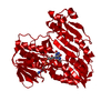 1d7yS S: Starting model for refinement |
|---|---|
| Similar structure data |


-Links
 PDBj
PDBj


- Assembly
| Deposited unit | 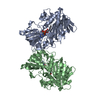
| ||||||||
|---|---|---|---|---|---|---|---|---|---|
| 1 | 
| ||||||||
| 2 | 
| ||||||||
| Unit cell |
| ||||||||
| Noncrystallographic symmetry (NCS) | NCS oper: (Code: given Matrix: (-0.99998, 0.00603, -0.00151), Vector : |
-Components
| #1: Protein | Mass: 57571.426 Da / Num. of mol.: 2 Source method: isolated from a genetically manipulated source Source: (gene. exp.) MUS MUSCULUS (house mouse) / Production host:  ESCHERICHIA COLI (E. coli) / References: UniProt: Q9Z0X1 ESCHERICHIA COLI (E. coli) / References: UniProt: Q9Z0X1#2: Chemical | Flavin adenine dinucleotide  Mass: 785.550 Da / Num. of mol.: 2 / Source method: obtained synthetically / Formula: C27H33N9O15P2 / Comment: FAD*YM Mass: 785.550 Da / Num. of mol.: 2 / Source method: obtained synthetically / Formula: C27H33N9O15P2 / Comment: FAD*YM#3: Water | ChemComp-HOH / | Water Mass: 18.015 Da / Num. of mol.: 440 / Source method: isolated from a natural source / Formula: H2O Mass: 18.015 Da / Num. of mol.: 440 / Source method: isolated from a natural source / Formula: H2O |
|---|
-Experimental details
-Experiment
| Experiment | Method: X-RAY DIFFRACTION / Number of used crystals: 1 |
|---|
- Sample preparation
| Crystal | Density Matthews: 2.3 Å3/Da / Density % sol: 43.5 % / Description: PHASE COMBINATION WITH MAD PHASES | ||||||||||||||||||||||||||||
|---|---|---|---|---|---|---|---|---|---|---|---|---|---|---|---|---|---|---|---|---|---|---|---|---|---|---|---|---|---|
| Crystal grow | pH: 7.75 Details: 18% PEG MONOMETHYL ETHER 5000, 100 MM MGCL2, 50 MM HEPES PH, pH 7.75 | ||||||||||||||||||||||||||||
| Crystal grow | *PLUS Method: vapor diffusion, hanging drop | ||||||||||||||||||||||||||||
| Components of the solutions | *PLUS
|
-Data collection
| Diffraction | Mean temperature: 100 K |
|---|---|
| Diffraction source | Source: SYNCHROTRON / Site: ESRF  / Beamline: ID14-4 / Wavelength: 0.93 / Beamline: ID14-4 / Wavelength: 0.93 |
| Detector | Detector: CCD / Date: Jun 15, 2000 |
| Radiation | Protocol: SINGLE WAVELENGTH / Monochromatic (M) / Laue (L): M / Scattering type: x-ray |
| Radiation wavelength | Wavelength: 0.93 Å / Relative weight: 1 |
| Reflection | Resolution: 2→20 Å / Num. obs: 62596 / % possible obs: 96.5 % / Redundancy: 2.9 % / Rmerge(I) obs: 0.11 / Net I/σ(I): 4.4 |
| Reflection shell | Resolution: 2→2.11 Å / Redundancy: 2.6 % / Rmerge(I) obs: 0.2 / Mean I/σ(I) obs: 2.5 / % possible all: 92.3 |
| Reflection | *PLUS Lowest resolution: 20 Å / % possible obs: 96.5 % / Rmerge(I) obs: 0.11 |
| Reflection shell | *PLUS % possible obs: 92.6 % / Rmerge(I) obs: 0.273 |
- Processing
| Software |
| ||||||||||||||||||||
|---|---|---|---|---|---|---|---|---|---|---|---|---|---|---|---|---|---|---|---|---|---|
| Refinement | Method to determine structure: MOLECULAR REPLACEMENT Starting model: 1D7Y Resolution: 2→15 Å / SU B: 5.03 / SU ML: 0.14 / Cross valid method: THROUGHOUT / ESU R Free: 0.183 Details: DISORDERED RESIDUES WERE MODELED USING ROTAMERS DATA BASE AND HAVE OCC=0.00 IN THE PDB
| ||||||||||||||||||||
| Refinement step | Cycle: LAST / Resolution: 2→15 Å
| ||||||||||||||||||||
| Refinement | *PLUS Num. reflection obs: 71317 / % reflection Rfree: 5 % / Rfactor obs: 0.216 / Rfactor Rfree: 0.257 / Rfactor Rwork: 0.216 | ||||||||||||||||||||
| Solvent computation | *PLUS | ||||||||||||||||||||
| Displacement parameters | *PLUS | ||||||||||||||||||||
| Refine LS restraints | *PLUS
| ||||||||||||||||||||
| LS refinement shell | *PLUS Rfactor Rfree: 0.339 / Rfactor Rwork: 0.24 / Rfactor obs: 0.24 |
