 Movie
Movie Controller


+ Open data

- Basic information
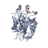
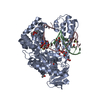
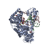
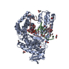
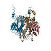
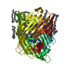
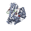
| Entry | Database: PDB / ID: 1gqf | ||||||
|---|---|---|---|---|---|---|---|
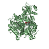
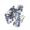
| Title | Crystal structure of human procaspase-7 | ||||||
 Components Components | Caspase-7 Caspase 7 Caspase 7 | ||||||
 Keywords Keywords | HYDROLASE / CASPASE-7 / APOPTOSIS / ZYMOGEN | ||||||
| Function / homology |  Function and homology informationcaspase-7 / lymphocyte apoptotic process / positive regulation of plasma membrane repair / cellular response to staurosporine / cysteine-type endopeptidase activity involved in execution phase of apoptosis / SMAC, XIAP-regulated apoptotic response / Activation of caspases through apoptosome-mediated cleavage / SMAC (DIABLO) binds to IAPs / SMAC(DIABLO)-mediated dissociation of IAP:caspase complexes / cysteine-type endopeptidase activity involved in apoptotic process ...caspase-7 / lymphocyte apoptotic process / positive regulation of plasma membrane repair / cellular response to staurosporine / cysteine-type endopeptidase activity involved in execution phase of apoptosis / SMAC, XIAP-regulated apoptotic response / Activation of caspases through apoptosome-mediated cleavage / SMAC (DIABLO) binds to IAPs / SMAC(DIABLO)-mediated dissociation of IAP:caspase complexes / cysteine-type endopeptidase activity involved in apoptotic process / fibroblast apoptotic process / execution phase of apoptosis / Apoptotic cleavage of cellular proteins / protein maturation / Caspase-mediated cleavage of cytoskeletal proteins / cysteine-type peptidase activity / response to UV / striated muscle cell differentiation / protein catabolic process / protein processing / heart development / peptidase activity / neuron apoptotic process / cellular response to lipopolysaccharide / aspartic-type endopeptidase activity / defense response to bacterium / cysteine-type endopeptidase activity / apoptotic process / proteolysis / extracellular space / RNA binding / nucleoplasm / nucleus / cytosol / cytoplasm Function and homology informationcaspase-7 / lymphocyte apoptotic process / positive regulation of plasma membrane repair / cellular response to staurosporine / cysteine-type endopeptidase activity involved in execution phase of apoptosis / SMAC, XIAP-regulated apoptotic response / Activation of caspases through apoptosome-mediated cleavage / SMAC (DIABLO) binds to IAPs / SMAC(DIABLO)-mediated dissociation of IAP:caspase complexes / cysteine-type endopeptidase activity involved in apoptotic process ...caspase-7 / lymphocyte apoptotic process / positive regulation of plasma membrane repair / cellular response to staurosporine / cysteine-type endopeptidase activity involved in execution phase of apoptosis / SMAC, XIAP-regulated apoptotic response / Activation of caspases through apoptosome-mediated cleavage / SMAC (DIABLO) binds to IAPs / SMAC(DIABLO)-mediated dissociation of IAP:caspase complexes / cysteine-type endopeptidase activity involved in apoptotic process / fibroblast apoptotic process / execution phase of apoptosis / Apoptotic cleavage of cellular proteins / protein maturation / Caspase-mediated cleavage of cytoskeletal proteins / cysteine-type peptidase activity / response to UV / striated muscle cell differentiation / protein catabolic process / protein processing / heart development / peptidase activity / neuron apoptotic process / cellular response to lipopolysaccharide / aspartic-type endopeptidase activity / defense response to bacterium / cysteine-type endopeptidase activity / apoptotic process / proteolysis / extracellular space / RNA binding / nucleoplasm / nucleus / cytosol / cytoplasmSimilarity search - Function | ||||||
| Biological species |  Homo sapiens (human) Homo sapiens (human) | ||||||
| Method | X-RAY DIFFRACTION / SYNCHROTRON / MOLECULAR REPLACEMENT / Resolution: 2.9 Å | ||||||
 Authors Authors | Riedl, S. / Bode, W. / Fuentes-Prior, P. | ||||||
 Citation Citation | Journal: Proc. Natl. Acad. Sci. U.S.A. / Year: 2001 Title: Structural basis for the activation of human procaspase-7. Authors: Riedl, S.J. / Fuentes-Prior, P. / Renatus, M. / Kairies, N. / Krapp, S. / Huber, R. / Salvesen, G.S. / Bode, W. | ||||||
| History |
|
- Structure visualization
| Structure viewer | Molecule: MolmilJmol/JSmol |
|---|
- Downloads & links
-Download
| PDBx/mmCIF format | 1gqf.cif.gz | 121.4 KB | Display | PDBx/mmCIF format |
|---|---|---|---|---|
| PDB format | pdb1gqf.ent.gz | 93.4 KB | Display | PDB format |
| PDBx/mmJSON format | 1gqf.json.gz | Tree view | PDBx/mmJSON format | |
| Others |  Other downloads Other downloads |
-Validation report
| Arichive directory | https://data.pdbj.org/pub/pdb/validation_reports/gq/1gqfftp://data.pdbj.org/pub/pdb/validation_reports/gq/1gqf | HTTPS FTP |
|---|
-Related structure data
| Related structure data | 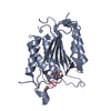 1f1jS S: Starting model for refinement |
|---|---|
| Similar structure data |

-Links
 PDBj
PDBj


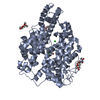
- Assembly
| Deposited unit | 
| ||||||||
|---|---|---|---|---|---|---|---|---|---|
| 1 |
| ||||||||
| Unit cell |
|
-Components
| #1: Protein | Caspase 7 / CASP-7 / Apoptotic protease Mch-3 / CMH-1 / ICE-like apoptotic protease 3 / ICE-LAP3 Mass: 30283.334 Da / Num. of mol.: 2 / Mutation: C187A Source method: isolated from a genetically manipulated source Source: (gene. exp.) Homo sapiens (human) / Gene: CASP7, MCH3 / Production host:  Escherichia coli (E. coli) / References: UniProt: P55210, caspase-7 Escherichia coli (E. coli) / References: UniProt: P55210, caspase-7#2: Chemical | ChemComp-SO4 / Sulfate  Mass: 96.063 Da / Num. of mol.: 5 / Source method: obtained synthetically / Formula: SO4 Mass: 96.063 Da / Num. of mol.: 5 / Source method: obtained synthetically / Formula: SO4#3: Water | ChemComp-HOH / | Water Mass: 18.015 Da / Num. of mol.: 20 / Source method: isolated from a natural source / Formula: H2O Mass: 18.015 Da / Num. of mol.: 20 / Source method: isolated from a natural source / Formula: H2O |
|---|
-Experimental details
-Experiment
| Experiment | Method: X-RAY DIFFRACTION / Number of used crystals: 1 |
|---|
- Sample preparation
| Crystal | Density Matthews: 3.55 Å3/Da / Density % sol: 60 % | |||||||||||||||||||||||||||||||||||||||||||||||||
|---|---|---|---|---|---|---|---|---|---|---|---|---|---|---|---|---|---|---|---|---|---|---|---|---|---|---|---|---|---|---|---|---|---|---|---|---|---|---|---|---|---|---|---|---|---|---|---|---|---|---|
| Crystal grow | Temperature: 293 K / Method: vapor diffusion / pH: 5.6 Details: 50 MM MES, PH 5.6, 10 MM MAGNESIUM CHLORIDE, 100 MM AMMONIUM SULFATE, 20% (W/V) PEG 8000 | |||||||||||||||||||||||||||||||||||||||||||||||||
| Crystal grow | *PLUS Temperature: 20 ℃ / Method: vapor diffusion, sitting drop / pH: 5.6 | |||||||||||||||||||||||||||||||||||||||||||||||||
| Components of the solutions | *PLUS
|
-Data collection
| Diffraction | Mean temperature: 100 K |
|---|---|
| Diffraction source | Source: SYNCHROTRON / Site: MPG/DESY, HAMBURG  / Beamline: BW6 / Wavelength: 1.05 / Beamline: BW6 / Wavelength: 1.05 |
| Detector | Type: MARRESEARCH / Detector: CCD / Date: Jul 23, 2001 |
| Radiation | Protocol: SINGLE WAVELENGTH / Monochromatic (M) / Laue (L): M / Scattering type: x-ray |
| Radiation wavelength | Wavelength: 1.05 Å / Relative weight: 1 |
| Reflection | Resolution: 2.9→59.76 Å / Num. obs: 19487 / % possible obs: 99.1 % / Observed criterion σ(I): 2 / Redundancy: 3.4 % / Biso Wilson estimate: 74.2 Å2 / Rmerge(I) obs: 0.068 |
| Reflection shell | Resolution: 2.9→3.06 Å / Redundancy: 3.1 % / Rmerge(I) obs: 0.386 / % possible all: 99.1 |
| Reflection | *PLUS Num. measured all: 180534 |
| Reflection shell | *PLUS % possible obs: 99.1 % |
- Processing
| Software |
| ||||||||||||||||||||||||||||||||||||||||||||||||||||||||||||
|---|---|---|---|---|---|---|---|---|---|---|---|---|---|---|---|---|---|---|---|---|---|---|---|---|---|---|---|---|---|---|---|---|---|---|---|---|---|---|---|---|---|---|---|---|---|---|---|---|---|---|---|---|---|---|---|---|---|---|---|---|---|
| Refinement | Method to determine structure: MOLECULAR REPLACEMENT Starting model: PDB ENTRY 1F1J Resolution: 2.9→21.73 Å / Rfactor Rfree error: 0.009 / Data cutoff high absF: 1166291.1 / Isotropic thermal model: RESTRAINED / Cross valid method: THROUGHOUT / σ(F): 0 Details: COMPETITION FOR THE LIMITED INTERMONOMER SPACE (THE "CENTRAL CAVITY") BY THE TWO INTERDOMAIN LINKERS MANIFESTS ITSELF IN POOR OR AMBIGOUS ELECTRON DENSITY FOR RESIDUES N-TERMINAL OF ILE321. ...Details: COMPETITION FOR THE LIMITED INTERMONOMER SPACE (THE "CENTRAL CAVITY") BY THE TWO INTERDOMAIN LINKERS MANIFESTS ITSELF IN POOR OR AMBIGOUS ELECTRON DENSITY FOR RESIDUES N-TERMINAL OF ILE321. ALTERNATE CONFORMATIONS, HOWEVER, ARE NOT SEPARABLE AT THE CURRENT RESOLUTION. ELECTRON DENSITY IS ALSO POOR OR MISSING FOR RESIDUES THR288 - LYS320 (THE INTERDOMAIN LINKER), GLY336 - GLY346 (THE SUBSTRATE BINDING LOOP), AS WELL AS FOR THE INSERTION LOOP GLU381-GLU382. RESIDUES N-TERMINAL OF PRO149 AND THE C-TERMINAL HISTIDINE TAG ARE FLEXIBLY DISORDERED.
| ||||||||||||||||||||||||||||||||||||||||||||||||||||||||||||
| Solvent computation | Solvent model: FLAT MODEL / Bsol: 210.532 Å2 / ksol: 0.237854 e/Å3 | ||||||||||||||||||||||||||||||||||||||||||||||||||||||||||||
| Displacement parameters | Biso mean: 80.7 Å2
| ||||||||||||||||||||||||||||||||||||||||||||||||||||||||||||
| Refine analyze |
| ||||||||||||||||||||||||||||||||||||||||||||||||||||||||||||
| Refinement step | Cycle: LAST / Resolution: 2.9→21.73 Å
| ||||||||||||||||||||||||||||||||||||||||||||||||||||||||||||
| Refine LS restraints |
| ||||||||||||||||||||||||||||||||||||||||||||||||||||||||||||
| LS refinement shell | Resolution: 2.9→3.08 Å / Rfactor Rfree error: 0.033 / Total num. of bins used: 6
| ||||||||||||||||||||||||||||||||||||||||||||||||||||||||||||
| Xplor file |
| ||||||||||||||||||||||||||||||||||||||||||||||||||||||||||||
| Software | *PLUS Name: CNS / Version: 1.1 / Classification: refinement | ||||||||||||||||||||||||||||||||||||||||||||||||||||||||||||
| Refinement | *PLUS Lowest resolution: 59.76 Å / % reflection Rfree: 5 % | ||||||||||||||||||||||||||||||||||||||||||||||||||||||||||||
| Solvent computation | *PLUS | ||||||||||||||||||||||||||||||||||||||||||||||||||||||||||||
| Displacement parameters | *PLUS | ||||||||||||||||||||||||||||||||||||||||||||||||||||||||||||
| Refine LS restraints | *PLUS
| ||||||||||||||||||||||||||||||||||||||||||||||||||||||||||||
| LS refinement shell | *PLUS Rfactor obs: 0.313 |
