 Movie
Movie Controller

[English] 日本語
 Yorodumi
- PDB-1f1j: CRYSTAL STRUCTURE OF CASPASE-7 IN COMPLEX WITH ACETYL-ASP-GLU-VAL... -
+ Open data

- Basic information
| Entry | Database: PDB / ID: 1f1j | ||||||
|---|---|---|---|---|---|---|---|
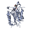
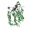
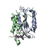
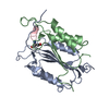
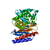
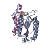
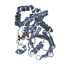
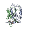
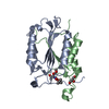
| Title | CRYSTAL STRUCTURE OF CASPASE-7 IN COMPLEX WITH ACETYL-ASP-GLU-VAL-ASP-CHO | ||||||
 Components Components |
| ||||||
 Keywords Keywords |  APOPTOSIS / HYDROLASE/HYDROLASE INHIBITOR / caspase-7 / cysteine protease / HYDROLASE / HYDROLASE-HYDROLASE INHIBITOR COMPLEX APOPTOSIS / HYDROLASE/HYDROLASE INHIBITOR / caspase-7 / cysteine protease / HYDROLASE / HYDROLASE-HYDROLASE INHIBITOR COMPLEX | ||||||
| Function / homology |  Function and homology informationcaspase-7 / lymphocyte apoptotic process / positive regulation of plasma membrane repair / cellular response to staurosporine / cysteine-type endopeptidase activity involved in execution phase of apoptosis / SMAC, XIAP-regulated apoptotic response / Activation of caspases through apoptosome-mediated cleavage / SMAC (DIABLO) binds to IAPs / SMAC(DIABLO)-mediated dissociation of IAP:caspase complexes / cysteine-type endopeptidase activity involved in apoptotic process ...caspase-7 / lymphocyte apoptotic process / positive regulation of plasma membrane repair / cellular response to staurosporine / cysteine-type endopeptidase activity involved in execution phase of apoptosis / SMAC, XIAP-regulated apoptotic response / Activation of caspases through apoptosome-mediated cleavage / SMAC (DIABLO) binds to IAPs / SMAC(DIABLO)-mediated dissociation of IAP:caspase complexes / cysteine-type endopeptidase activity involved in apoptotic process / fibroblast apoptotic process / execution phase of apoptosis / Apoptotic cleavage of cellular proteins / protein maturation / Caspase-mediated cleavage of cytoskeletal proteins / cysteine-type peptidase activity / response to UV / striated muscle cell differentiation / protein catabolic process / protein processing / heart development / peptidase activity / neuron apoptotic process / cellular response to lipopolysaccharide / aspartic-type endopeptidase activity / defense response to bacterium / cysteine-type endopeptidase activity / apoptotic process / proteolysis / extracellular space / RNA binding / nucleoplasm / nucleus / cytosol / cytoplasm Function and homology informationcaspase-7 / lymphocyte apoptotic process / positive regulation of plasma membrane repair / cellular response to staurosporine / cysteine-type endopeptidase activity involved in execution phase of apoptosis / SMAC, XIAP-regulated apoptotic response / Activation of caspases through apoptosome-mediated cleavage / SMAC (DIABLO) binds to IAPs / SMAC(DIABLO)-mediated dissociation of IAP:caspase complexes / cysteine-type endopeptidase activity involved in apoptotic process ...caspase-7 / lymphocyte apoptotic process / positive regulation of plasma membrane repair / cellular response to staurosporine / cysteine-type endopeptidase activity involved in execution phase of apoptosis / SMAC, XIAP-regulated apoptotic response / Activation of caspases through apoptosome-mediated cleavage / SMAC (DIABLO) binds to IAPs / SMAC(DIABLO)-mediated dissociation of IAP:caspase complexes / cysteine-type endopeptidase activity involved in apoptotic process / fibroblast apoptotic process / execution phase of apoptosis / Apoptotic cleavage of cellular proteins / protein maturation / Caspase-mediated cleavage of cytoskeletal proteins / cysteine-type peptidase activity / response to UV / striated muscle cell differentiation / protein catabolic process / protein processing / heart development / peptidase activity / neuron apoptotic process / cellular response to lipopolysaccharide / aspartic-type endopeptidase activity / defense response to bacterium / cysteine-type endopeptidase activity / apoptotic process / proteolysis / extracellular space / RNA binding / nucleoplasm / nucleus / cytosol / cytoplasmSimilarity search - Function | ||||||
| Biological species |  Homo sapiens (human) Homo sapiens (human) | ||||||
| Method | X-RAY DIFFRACTION / SYNCHROTRON / Resolution: 2.35 Å | ||||||
 Authors Authors | Wei, Y. / Charifson, P.S. | ||||||
 Citation Citation | Journal: Chem.Biol. / Year: 2000 Title: The structures of caspases-1, -3, -7 and -8 reveal the basis for substrate and inhibitor selectivity. Authors: Wei, Y. / Fox, T. / Chambers, S.P. / Sintchak, J. / Coll, J.T. / Golec, J.M. / Swenson, L. / Wilson, K.P. / Charifson, P.S. | ||||||
| History |
|
- Structure visualization
| Structure viewer | Molecule: MolmilJmol/JSmol |
|---|
- Downloads & links
-Download
| PDBx/mmCIF format | 1f1j.cif.gz | 113.9 KB | Display | PDBx/mmCIF format |
|---|---|---|---|---|
| PDB format | pdb1f1j.ent.gz | 91.2 KB | Display | PDB format |
| PDBx/mmJSON format | 1f1j.json.gz | Tree view | PDBx/mmJSON format | |
| Others |  Other downloads Other downloads |
-Validation report
| Arichive directory | https://data.pdbj.org/pub/pdb/validation_reports/f1/1f1jftp://data.pdbj.org/pub/pdb/validation_reports/f1/1f1j | HTTPS FTP |
|---|
-Related structure data
| Similar structure data |
|---|

-Links
 PDBj
PDBj


- Assembly
| Deposited unit | 
| ||||||||
|---|---|---|---|---|---|---|---|---|---|
| 1 | 
| ||||||||
| 2 | 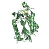
| ||||||||
| 3 | 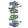
| ||||||||
| 4 |
| ||||||||
| 5 | 
| ||||||||
| Unit cell |
|
-Components
| #1: Protein | Mass: 34550.031 Da / Num. of mol.: 2 / Fragment: P20/P10 CATALYTIC DOMAIN Source method: isolated from a genetically manipulated source Source: (gene. exp.) Homo sapiens (human) / Production host:  Escherichia coli (E. coli) Escherichia coli (E. coli)References: UniProt: P55210, Hydrolases; Acting on peptide bonds (peptidases); Cysteine endopeptidases#2: Protein/peptide | 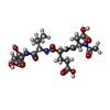  Type: Peptide-like / Class: Inhibitor / Mass: 488.489 Da / Num. of mol.: 2 / Source method: obtained synthetically / Details: this sequence was chemically synthesized / References: Ac-Asp-Glu-Val-Asp-Aldehyde Type: Peptide-like / Class: Inhibitor / Mass: 488.489 Da / Num. of mol.: 2 / Source method: obtained synthetically / Details: this sequence was chemically synthesized / References: Ac-Asp-Glu-Val-Asp-Aldehyde#3: Chemical | Sulfate  Mass: 96.063 Da / Num. of mol.: 2 / Source method: obtained synthetically / Formula: SO4 Mass: 96.063 Da / Num. of mol.: 2 / Source method: obtained synthetically / Formula: SO4#4: Water | ChemComp-HOH / | Water Mass: 18.015 Da / Num. of mol.: 397 / Source method: isolated from a natural source / Formula: H2O Mass: 18.015 Da / Num. of mol.: 397 / Source method: isolated from a natural source / Formula: H2OCompound details | THERE IS A COVALENT BOND BETWEEN ATOM SG OF CYS186A AND ATOM C OF ASA705C, AND SAME BOND BETWEEN ...THERE IS A COVALENT BOND BETWEEN ATOM SG OF CYS186A AND ATOM C OF ASA705C, AND SAME BOND BETWEEN ATOM SG OF CYS486B AND ATOM C OF ASA805D, FORMING THIOHEMIAC | |
|---|
-Experimental details
-Experiment
| Experiment | Method: X-RAY DIFFRACTION / Number of used crystals: 1 |
|---|
- Sample preparation
| Crystal | Density Matthews: 2.98 Å3/Da / Density % sol: 58.73 % | ||||||||||||||||||||||||||||||||||||||||||||||||||||||
|---|---|---|---|---|---|---|---|---|---|---|---|---|---|---|---|---|---|---|---|---|---|---|---|---|---|---|---|---|---|---|---|---|---|---|---|---|---|---|---|---|---|---|---|---|---|---|---|---|---|---|---|---|---|---|---|
| Crystal grow | Temperature: 277 K / Method: vapor diffusion, hanging drop / pH: 7 Details: 30% PEG 4000, 0.1 M Na-citrate, 0.2 M ammonium acetate, pH 7.0, VAPOR DIFFUSION, HANGING DROP, temperature 277K | ||||||||||||||||||||||||||||||||||||||||||||||||||||||
| Crystal grow | *PLUS Method: vapor diffusion / Details: used microseeding | ||||||||||||||||||||||||||||||||||||||||||||||||||||||
| Components of the solutions | *PLUS
|
-Data collection
| Diffraction | Mean temperature: 128 K |
|---|---|
| Diffraction source | Source: SYNCHROTRON / Site: NSLS  / Beamline: X25 / Wavelength: 1.1 / Beamline: X25 / Wavelength: 1.1 |
| Detector | Type: BRANDEIS - B4 / Detector: CCD / Date: Sep 20, 1998 |
| Radiation | Protocol: SINGLE WAVELENGTH / Monochromatic (M) / Laue (L): M / Scattering type: x-ray |
| Radiation wavelength | Wavelength: 1.1 Å / Relative weight: 1 |
| Reflection | Resolution: 2.35→20 Å / Num. all: 35568 / Num. obs: 34359 / % possible obs: 96.6 % / Observed criterion σ(F): 2 / Observed criterion σ(I): 2 / Redundancy: 4.1 % / Biso Wilson estimate: 26 Å2 / Rmerge(I) obs: 0.083 / Net I/σ(I): 16.5 |
| Reflection shell | Resolution: 2.35→2.42 Å / Redundancy: 2.7 % / Rmerge(I) obs: 0.41 / % possible all: 92.3 |
| Reflection | *PLUS Num. measured all: 139781 |
- Processing
| Software |
| ||||||||||||||||||||
|---|---|---|---|---|---|---|---|---|---|---|---|---|---|---|---|---|---|---|---|---|---|
| Refinement | Resolution: 2.35→7.5 Å / σ(F): 2.5 / σ(I): 1.6 / Stereochemistry target values: Engh & Huber
| ||||||||||||||||||||
| Refinement step | Cycle: LAST / Resolution: 2.35→7.5 Å
| ||||||||||||||||||||
| Refine LS restraints |
| ||||||||||||||||||||
| Software | *PLUS Name: X-PLOR / Version: 3.843 / Classification: refinement | ||||||||||||||||||||
| Refinement | *PLUS Lowest resolution: 7.5 Å / σ(F): 2.5 / % reflection Rfree: 5 % | ||||||||||||||||||||
| Solvent computation | *PLUS | ||||||||||||||||||||
| Displacement parameters | *PLUS | ||||||||||||||||||||
| Refine LS restraints | *PLUS
|
