 Movie
Movie Controller

[English] 日本語
 Yorodumi
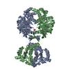
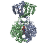
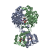
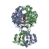
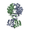
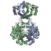
- PDB-1g8m: CRYSTAL STRUCTURE OF AVIAN ATIC, A BIFUNCTIONAL TRANSFORMYLASE AN... -
+ Open data

- Basic information
| Entry | Database: PDB / ID: 1g8m | ||||||
|---|---|---|---|---|---|---|---|
| Title | CRYSTAL STRUCTURE OF AVIAN ATIC, A BIFUNCTIONAL TRANSFORMYLASE AND CYCLOHYDROLASE ENZYME IN PURINE BIOSYNTHESIS AT 1.75 ANG. RESOLUTION | ||||||
 Components Components | AICAR TRANSFORMYLASE-IMP CYCLOHYDROLASE | ||||||
 Keywords Keywords |  transferase / hydrolase / Homodimer / 2 functional domains / IMPCH domain = alpha/beta/alpha / AICAR Tfase = 2 alpha/beta/alpha domains / 1 alpha + beta domain transferase / hydrolase / Homodimer / 2 functional domains / IMPCH domain = alpha/beta/alpha / AICAR Tfase = 2 alpha/beta/alpha domains / 1 alpha + beta domain | ||||||
| Function / homology |  Function and homology information Function and homology informationDe novo synthesis of IMP / phosphoribosylaminoimidazolecarboxamide formyltransferase / phosphoribosylaminoimidazolecarboxamide formyltransferase activity / IMP cyclohydrolase / IMP cyclohydrolase activity / 'de novo' IMP biosynthetic process / protein homodimerization activity / cytosolSimilarity search - Function | ||||||
| Biological species |  Gallus gallus (chicken) Gallus gallus (chicken) | ||||||
| Method | X-RAY DIFFRACTION / SYNCHROTRON / MAD / Resolution: 1.75 Å | ||||||
 Authors Authors | Greasley, S.E. / Horton, P. / Beardsley, G.P. / Benkovic, S.J. / Wilson, I.A. | ||||||
 Citation Citation | Journal: Nat.Struct.Biol. / Year: 2001 Title: Crystal structure of a bifunctional transformylase and cyclohydrolase enzyme in purine biosynthesis. Authors: Greasley, S.E. / Horton, P. / Ramcharan, J. / Beardsley, G.P. / Benkovic, S.J. / Wilson, I.A. | ||||||
| History |
|
- Structure visualization
| Structure viewer | Molecule: MolmilJmol/JSmol |
|---|
- Downloads & links
-Download
| PDBx/mmCIF format | 1g8m.cif.gz | 246.7 KB | Display | PDBx/mmCIF format |
|---|---|---|---|---|
| PDB format | pdb1g8m.ent.gz | 204.2 KB | Display | PDB format |
| PDBx/mmJSON format | 1g8m.json.gz | Tree view | PDBx/mmJSON format | |
| Others |  Other downloads Other downloads |
-Validation report
| Arichive directory | https://data.pdbj.org/pub/pdb/validation_reports/g8/1g8mftp://data.pdbj.org/pub/pdb/validation_reports/g8/1g8m | HTTPS FTP |
|---|
-Related structure data
| Similar structure data |
|---|


-Links
 PDBj
PDBj


- Assembly
| Deposited unit | 
| ||||||||||
|---|---|---|---|---|---|---|---|---|---|---|---|
| 1 |
| ||||||||||
| Unit cell |
|
-Components
| #1: Protein | Mass: 64966.656 Da / Num. of mol.: 2 Fragment: AMINOIMIDAZOLE CARBOXAMIDE RIBONUCLEOTIDE TRANSFORMYLASE - INOSINE MONOPHOSPHATE CYCLOHYDROLASE Mutation: MET REPLACED BY SE-MET (MSE) Source method: isolated from a genetically manipulated source Source: (gene. exp.) Gallus gallus (chicken) / Gene: PURH / Plasmid: PET 28A / Production host:  Escherichia coli (E. coli) / Strain (production host): B834(DE3) Escherichia coli (E. coli) / Strain (production host): B834(DE3)References: UniProt: P31335, phosphoribosylaminoimidazolecarboxamide formyltransferase, IMP cyclohydrolase#2: Chemical |   Mass: 39.098 Da / Num. of mol.: 2 / Source method: obtained synthetically / Formula: K Mass: 39.098 Da / Num. of mol.: 2 / Source method: obtained synthetically / Formula: K#3: Chemical | ChemComp-G / | Guanosine monophosphate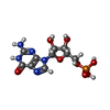  Type: RNA linking / Mass: 363.221 Da / Num. of mol.: 1 / Source method: obtained synthetically / Formula: C10H14N5O8P Type: RNA linking / Mass: 363.221 Da / Num. of mol.: 1 / Source method: obtained synthetically / Formula: C10H14N5O8P#4: Water | ChemComp-HOH / | Water Mass: 18.015 Da / Num. of mol.: 744 / Source method: isolated from a natural source / Formula: H2O Mass: 18.015 Da / Num. of mol.: 744 / Source method: isolated from a natural source / Formula: H2O |
|---|
-Experimental details
-Experiment
| Experiment | Method: X-RAY DIFFRACTION / Number of used crystals: 1 |
|---|
- Sample preparation
| Crystal | Density Matthews: 2.61 Å3/Da / Density % sol: 52.92 % | ||||||||||||||||||||||||||||||||||||||||||||||||||||||||||||
|---|---|---|---|---|---|---|---|---|---|---|---|---|---|---|---|---|---|---|---|---|---|---|---|---|---|---|---|---|---|---|---|---|---|---|---|---|---|---|---|---|---|---|---|---|---|---|---|---|---|---|---|---|---|---|---|---|---|---|---|---|---|
| Crystal grow | Temperature: 295 K / Method: vapor diffusion, sitting drop / pH: 7.2 Details: PEG 8000, imidazole-HCl and DTT, pH 7.2, VAPOR DIFFUSION, SITTING DROP at 295K | ||||||||||||||||||||||||||||||||||||||||||||||||||||||||||||
| Crystal | *PLUS Density % sol: 53 % | ||||||||||||||||||||||||||||||||||||||||||||||||||||||||||||
| Crystal grow | *PLUS Temperature: 22 ℃ / pH: 7.4 / Method: vapor diffusion | ||||||||||||||||||||||||||||||||||||||||||||||||||||||||||||
| Components of the solutions | *PLUS
|
-Data collection
| Diffraction | Mean temperature: 93 K | ||||||||||||
|---|---|---|---|---|---|---|---|---|---|---|---|---|---|
| Diffraction source | Source: SYNCHROTRON / Site: ALS  / Beamline: 5.0.2 / Wavelength: 0.9799 / Wavelength: 0.9799, 0.9801, 0.9648 / Beamline: 5.0.2 / Wavelength: 0.9799 / Wavelength: 0.9799, 0.9801, 0.9648 | ||||||||||||
| Detector | Type: ADSC QUANTUM 4 / Detector: CCD / Date: Feb 3, 1999 | ||||||||||||
| Radiation | Monochromator: Double crystal / Protocol: MAD / Monochromatic (M) / Laue (L): M / Scattering type: x-ray | ||||||||||||
| Radiation wavelength |
| ||||||||||||
| Reflection | Resolution: 1.75→50 Å / Num. all: 229603 / Num. obs: 229603 / % possible obs: 87.9 % / Observed criterion σ(F): 0 / Observed criterion σ(I): -3 / Redundancy: 2 % / Biso Wilson estimate: 15.9 Å2 / Rmerge(I) obs: 0.058 / Net I/σ(I): 15.8 | ||||||||||||
| Reflection shell | Resolution: 1.75→1.78 Å / Redundancy: 2 % / Rmerge(I) obs: 0.475 / Mean I/σ(I) obs: 2.4 / % possible all: 47.3 | ||||||||||||
| Reflection | *PLUS Num. measured all: 460370 | ||||||||||||
| Reflection shell | *PLUS % possible obs: 47.3 % |
- Processing
| Software |
| ||||||||||||||||||||||||||||||||||||
|---|---|---|---|---|---|---|---|---|---|---|---|---|---|---|---|---|---|---|---|---|---|---|---|---|---|---|---|---|---|---|---|---|---|---|---|---|---|
| Refinement | Method to determine structure: MAD / Resolution: 1.75→50 Å / Rfactor Rfree error: 0.002 / Data cutoff high absF: 1962794.87 / Data cutoff low absF: 0 / Isotropic thermal model: RESTRAINED / Cross valid method: THROUGHOUT / σ(F): 0 / Stereochemistry target values: Engh & Huber
| ||||||||||||||||||||||||||||||||||||
| Solvent computation | Solvent model: FLAT MODEL / Bsol: 39.74 Å2 / ksol: 0.348 e/Å3 | ||||||||||||||||||||||||||||||||||||
| Displacement parameters | Biso mean: 19.2 Å2
| ||||||||||||||||||||||||||||||||||||
| Refine analyze |
| ||||||||||||||||||||||||||||||||||||
| Refinement step | Cycle: LAST / Resolution: 1.75→50 Å
| ||||||||||||||||||||||||||||||||||||
| Refine LS restraints |
| ||||||||||||||||||||||||||||||||||||
| LS refinement shell | Resolution: 1.75→1.86 Å / Rfactor Rfree error: 0.008 / Total num. of bins used: 6
| ||||||||||||||||||||||||||||||||||||
| Xplor file |
| ||||||||||||||||||||||||||||||||||||
| Software | *PLUS Name: CNS / Version: 0.9 / Classification: refinement | ||||||||||||||||||||||||||||||||||||
| Refine LS restraints | *PLUS
|
