 Movie
Movie Controller


+ Open data

- Basic information
| Entry | Database: PDB / ID: 1fsl | ||||||
|---|---|---|---|---|---|---|---|
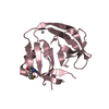
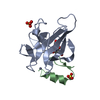
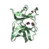
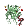
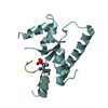
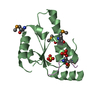
| Title | FERRIC SOYBEAN LEGHEMOGLOBIN COMPLEXED WITH NICOTINATE | ||||||
 Components Components | LEGHEMOGLOBIN A | ||||||
 Keywords Keywords |  OXYGEN TRANSPORT / HEME / RESPIRATORY PROTEIN / NITROGEN FIXATION / MULTIGENE FAMILY OXYGEN TRANSPORT / HEME / RESPIRATORY PROTEIN / NITROGEN FIXATION / MULTIGENE FAMILY | ||||||
| Function / homology |  Function and homology information Function and homology informationresponse to nitrogen compound / nodulation / response to hydrogen sulfide / response to cobalt ion / oxygen carrier activity / oxygen binding / heme binding / metal ion bindingSimilarity search - Function | ||||||
| Biological species |  Glycine max (soybean) Glycine max (soybean) | ||||||
| Method | X-RAY DIFFRACTION / MIR / Resolution: 2.3 Å | ||||||
 Authors Authors | Ellis, P.J. / Guss, J.M. / Freeman, H.C. | ||||||
 Citation Citation | Journal: Acta Crystallogr.,Sect.D / Year: 1997 Title: Structure of ferric soybean leghemoglobin a nicotinate at 2.3 A resolution. Authors: Ellis, P.J. / Appleby, C.A. / Guss, J.M. / Hunter, W.N. / Ollis, D.L. / Freeman, H.C. #1: Journal: Aust.J.Chem. / Year: 1983Title: Crystal Structure of Soybean Ferric Leghaemoglobin a Nicotinate at a Resolution of 3.3 Angstrom Authors: Ollis, D.L. / Appleby, C.A. / Colman, P.M. / Cutten, A.E. / Guss, J.M. / Venkatappa, M.P. / Freeman, H.C. | ||||||
| History |
|
- Structure visualization
| Structure viewer | Molecule: MolmilJmol/JSmol |
|---|
- Downloads & links
-Download
| PDBx/mmCIF format | 1fsl.cif.gz | 117.4 KB | Display | PDBx/mmCIF format |
|---|---|---|---|---|
| PDB format | pdb1fsl.ent.gz | 94.5 KB | Display | PDB format |
| PDBx/mmJSON format | 1fsl.json.gz | Tree view | PDBx/mmJSON format | |
| Others |  Other downloads Other downloads |
-Validation report
| Arichive directory | https://data.pdbj.org/pub/pdb/validation_reports/fs/1fslftp://data.pdbj.org/pub/pdb/validation_reports/fs/1fsl | HTTPS FTP |
|---|
-Related structure data
| Similar structure data |
|---|


-Links
 PDBj
PDBj


- Assembly
| Deposited unit | 
| ||||||||
|---|---|---|---|---|---|---|---|---|---|
| 1 | 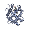
| ||||||||
| 2 | 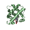
| ||||||||
| Unit cell |
| ||||||||
| Noncrystallographic symmetry (NCS) | NCS oper: (Code: given Matrix: (-0.461976, 0.030242, 0.886377), Vector : |
-Components
| #1: Protein | Mass: 15260.336 Da / Num. of mol.: 2 / Source method: isolated from a natural source / Details: FE(III), PH 6.0 / Source: (natural) Glycine max (soybean) / Organ: ROOT NODULE / References: UniProt: P02238#2: Chemical | Heme B  Mass: 616.487 Da / Num. of mol.: 2 / Source method: obtained synthetically / Formula: C34H32FeN4O4 Mass: 616.487 Da / Num. of mol.: 2 / Source method: obtained synthetically / Formula: C34H32FeN4O4#3: Chemical | Niacin  Mass: 123.109 Da / Num. of mol.: 2 / Source method: obtained synthetically / Formula: C6H5NO2 Mass: 123.109 Da / Num. of mol.: 2 / Source method: obtained synthetically / Formula: C6H5NO2#4: Water | ChemComp-HOH / | Water Mass: 18.015 Da / Num. of mol.: 70 / Source method: isolated from a natural source / Formula: H2O Mass: 18.015 Da / Num. of mol.: 70 / Source method: isolated from a natural source / Formula: H2O |
|---|
-Experimental details
-Experiment
| Experiment | Method: X-RAY DIFFRACTION / Number of used crystals: 6 |
|---|
- Sample preparation
| Crystal | Density Matthews: 2.65 Å3/Da / Density % sol: 53.62 % |
|---|---|
| Crystal grow | pH: 6 / Details: pH 6.0 |
| Crystal grow | *PLUS Method: other / Details: Ollis, D.L., (1983) Aust. J. Chem., 36, 451. |
-Data collection
| Diffraction | Mean temperature: 295 K |
|---|---|
| Diffraction source | Source: SEALED TUBE / Type: OTHER / Wavelength: 1.5418 |
| Detector | Type: ENRAF-NONIUS FAST / Detector: DIFFRACTOMETER / Date: 1981 / Details: COLLIMATOR |
| Radiation | Monochromator: NI FILTER / Monochromatic (M) / Laue (L): M / Scattering type: x-ray |
| Radiation wavelength | Wavelength: 1.5418 Å / Relative weight: 1 |
| Reflection | Resolution: 2.2→8 Å / Num. obs: 14153 / Observed criterion σ(I): 0 / Redundancy: 1.3 % / Rmerge(I) obs: 0.061 |
| Reflection | *PLUS Num. measured all: 18416 |
- Processing
| Software |
| ||||||||||||||||||||||||||||||||||||||||||||||||||||||||||||||||||||||||||||||||||||
|---|---|---|---|---|---|---|---|---|---|---|---|---|---|---|---|---|---|---|---|---|---|---|---|---|---|---|---|---|---|---|---|---|---|---|---|---|---|---|---|---|---|---|---|---|---|---|---|---|---|---|---|---|---|---|---|---|---|---|---|---|---|---|---|---|---|---|---|---|---|---|---|---|---|---|---|---|---|---|---|---|---|---|---|---|---|
| Refinement | Method to determine structure: MIR / Resolution: 2.3→6 Å / σ(F): 2
| ||||||||||||||||||||||||||||||||||||||||||||||||||||||||||||||||||||||||||||||||||||
| Displacement parameters | Biso mean: 12.3 Å2 | ||||||||||||||||||||||||||||||||||||||||||||||||||||||||||||||||||||||||||||||||||||
| Refine analyze | Luzzati coordinate error obs: 0.18 Å | ||||||||||||||||||||||||||||||||||||||||||||||||||||||||||||||||||||||||||||||||||||
| Refinement step | Cycle: LAST / Resolution: 2.3→6 Å
| ||||||||||||||||||||||||||||||||||||||||||||||||||||||||||||||||||||||||||||||||||||
| Refine LS restraints |
| ||||||||||||||||||||||||||||||||||||||||||||||||||||||||||||||||||||||||||||||||||||
| Software | *PLUS Name: PROFFT / Classification: refinement | ||||||||||||||||||||||||||||||||||||||||||||||||||||||||||||||||||||||||||||||||||||
| Refinement | *PLUS Rfactor obs: 0.158 | ||||||||||||||||||||||||||||||||||||||||||||||||||||||||||||||||||||||||||||||||||||
| Solvent computation | *PLUS | ||||||||||||||||||||||||||||||||||||||||||||||||||||||||||||||||||||||||||||||||||||
| Displacement parameters | *PLUS |
