 Movie
Movie Controller

[English] 日本語
 Yorodumi
- PDB-1fp3: CRYSTAL STRUCTURE OF N-ACYL-D-GLUCOSAMINE 2-EPIMERASE FROM PORCIN... -
+ Open data

- Basic information
| Entry | Database: PDB / ID: 1fp3 | ||||||
|---|---|---|---|---|---|---|---|
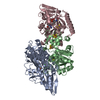
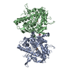
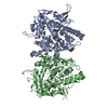
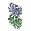
| Title | CRYSTAL STRUCTURE OF N-ACYL-D-GLUCOSAMINE 2-EPIMERASE FROM PORCINE KIDNEY | ||||||
 Components Components | N-ACYL-D-GLUCOSAMINE 2-EPIMERASE | ||||||
 Keywords Keywords |  ISOMERASE / ALPHA/ALPHA-BARREL / N-acyl-D-glucosamine 2-epimerase ISOMERASE / ALPHA/ALPHA-BARREL / N-acyl-D-glucosamine 2-epimerase | ||||||
| Function / homology |  Function and homology informationN-acylglucosamine 2-epimerase / N-acylglucosamine 2-epimerase activity / N-acetylmannosamine metabolic process / peptidase inhibitor activity / N-acetylglucosamine metabolic process / N-acetylneuraminate catabolic process Function and homology informationN-acylglucosamine 2-epimerase / N-acylglucosamine 2-epimerase activity / N-acetylmannosamine metabolic process / peptidase inhibitor activity / N-acetylglucosamine metabolic process / N-acetylneuraminate catabolic processSimilarity search - Function | ||||||
| Biological species |  Sus scrofa (pig) Sus scrofa (pig) | ||||||
| Method | X-RAY DIFFRACTION / Resolution: 2 Å | ||||||
 Authors Authors | Itoh, T. / Mikami, B. / Maru, I. / Ohta, Y. / Hashimoto, W. / Murata, K. | ||||||
 Citation Citation | Journal: J.Mol.Biol. / Year: 2000 Title: Crystal structure of N-acyl-D-glucosamine 2-epimerase from porcine kidney at 2.0 A resolution. Authors: Itoh, T. / Mikami, B. / Maru, I. / Ohta, Y. / Hashimoto, W. / Murata, K. | ||||||
| History |
|
- Structure visualization
| Structure viewer | Molecule: MolmilJmol/JSmol |
|---|
- Downloads & links
-Download
| PDBx/mmCIF format | 1fp3.cif.gz | 165.6 KB | Display | PDBx/mmCIF format |
|---|---|---|---|---|
| PDB format | pdb1fp3.ent.gz | 134.7 KB | Display | PDB format |
| PDBx/mmJSON format | 1fp3.json.gz | Tree view | PDBx/mmJSON format | |
| Others |  Other downloads Other downloads |
-Validation report
| Arichive directory | https://data.pdbj.org/pub/pdb/validation_reports/fp/1fp3ftp://data.pdbj.org/pub/pdb/validation_reports/fp/1fp3 | HTTPS FTP |
|---|
-Related structure data
| Similar structure data |
|---|


-Links
 PDBj
PDBj
- Assembly
| Deposited unit | 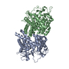
| ||||||||
|---|---|---|---|---|---|---|---|---|---|
| 1 |
| ||||||||
| 2 | 
| ||||||||
| Unit cell |
| ||||||||
| Details | The biological assembly is a dimer related by the two-fold axis. |
-Components
| #1: Protein | Mass: 46465.059 Da / Num. of mol.: 2 Source method: isolated from a genetically manipulated source Source: (gene. exp.) Sus scrofa (pig) / Tissue: KIDNEY / Plasmid: PEP114 / Production host:  Escherichia coli (E. coli) / References: UniProt: P17560, N-acylglucosamine 2-epimerase Escherichia coli (E. coli) / References: UniProt: P17560, N-acylglucosamine 2-epimerase#2: Water | ChemComp-HOH / | Water Mass: 18.015 Da / Num. of mol.: 145 / Source method: isolated from a natural source / Formula: H2O / References: N-acylglucosamine 2-epimerase Mass: 18.015 Da / Num. of mol.: 145 / Source method: isolated from a natural source / Formula: H2O / References: N-acylglucosamine 2-epimerase |
|---|
-Experimental details
-Experiment
| Experiment | Method: X-RAY DIFFRACTION / Number of used crystals: 1 |
|---|
- Sample preparation
| Crystal | Density Matthews: 2.06 Å3/Da / Density % sol: 40.16 % | ||||||||||||||||||||||||||||||||||||||||||||||||||||||
|---|---|---|---|---|---|---|---|---|---|---|---|---|---|---|---|---|---|---|---|---|---|---|---|---|---|---|---|---|---|---|---|---|---|---|---|---|---|---|---|---|---|---|---|---|---|---|---|---|---|---|---|---|---|---|---|
| Crystal grow | Temperature: 293 K / Method: vapor diffusion, hanging drop / pH: 5 Details: PEG 6000, sodium citrate, ammonium acetate, pH 5.0, VAPOR DIFFUSION, HANGING DROP, temperature 293.0K | ||||||||||||||||||||||||||||||||||||||||||||||||||||||
| Crystal | *PLUS Density % sol: 58 % | ||||||||||||||||||||||||||||||||||||||||||||||||||||||
| Crystal grow | *PLUS Temperature: 20 ℃ / Details: Maru, I., (1996) J. Biochem., 120, 481. | ||||||||||||||||||||||||||||||||||||||||||||||||||||||
| Components of the solutions | *PLUS
|
-Data collection
| Diffraction | Mean temperature: 293 K |
|---|---|
| Diffraction source | Source: ROTATING ANODE / Type: MACSCIENCE / Wavelength: 1.5418 |
| Detector | Type: SIEMENS HI-STAR / Detector: AREA DETECTOR / Date: Apr 19, 1996 |
| Radiation | Protocol: SINGLE WAVELENGTH / Monochromatic (M) / Laue (L): M / Scattering type: x-ray |
| Radiation wavelength | Wavelength: 1.5418 Å / Relative weight: 1 |
| Reflection | Resolution: 1.92→25 Å / Num. all: 49059 / Num. obs: 39143 / % possible obs: 82.1 % / Observed criterion σ(I): 1 / Redundancy: 2.5 % / Biso Wilson estimate: 10 Å2 / Rmerge(I) obs: 0.056 |
| Reflection shell | Resolution: 1.92→1.98 Å / Redundancy: 2.54 % / Rmerge(I) obs: 0.056 / Num. unique all: 1270 / % possible all: 82.1 |
| Reflection | *PLUS Num. measured all: 124509 |
- Processing
| Software |
| ||||||||||||||||||||||||||||||||||||||||||||
|---|---|---|---|---|---|---|---|---|---|---|---|---|---|---|---|---|---|---|---|---|---|---|---|---|---|---|---|---|---|---|---|---|---|---|---|---|---|---|---|---|---|---|---|---|---|
| Refinement | Resolution: 2→14.97 Å / Rfactor Rfree error: 0.004 / Data cutoff high absF: 667176.13 / Data cutoff low absF: 0 / Isotropic thermal model: RESTRAINED / Cross valid method: THROUGHOUT / σ(F): 2 / Stereochemistry target values: Engh & Huber
| ||||||||||||||||||||||||||||||||||||||||||||
| Solvent computation | Solvent model: FLAT MODEL / Bsol: 40.37 Å2 / ksol: 0.342 e/Å3 | ||||||||||||||||||||||||||||||||||||||||||||
| Displacement parameters | Biso mean: 25.5 Å2
| ||||||||||||||||||||||||||||||||||||||||||||
| Refine analyze |
| ||||||||||||||||||||||||||||||||||||||||||||
| Refinement step | Cycle: LAST / Resolution: 2→14.97 Å
| ||||||||||||||||||||||||||||||||||||||||||||
| Refine LS restraints |
| ||||||||||||||||||||||||||||||||||||||||||||
| LS refinement shell | Resolution: 2→2.12 Å / Rfactor Rfree error: 0.014 / Total num. of bins used: 6
| ||||||||||||||||||||||||||||||||||||||||||||
| Xplor file |
| ||||||||||||||||||||||||||||||||||||||||||||
| Software | *PLUS Name: X-PLOR / Version: 3.851 / Classification: refinement | ||||||||||||||||||||||||||||||||||||||||||||
| Refinement | *PLUS σ(F): 2 / % reflection Rfree: 10 % | ||||||||||||||||||||||||||||||||||||||||||||
| Solvent computation | *PLUS | ||||||||||||||||||||||||||||||||||||||||||||
| Displacement parameters | *PLUS Biso mean: 25.5 Å2 | ||||||||||||||||||||||||||||||||||||||||||||
| Refine LS restraints | *PLUS
| ||||||||||||||||||||||||||||||||||||||||||||
| LS refinement shell | *PLUS Rfactor Rfree: 0.294 / % reflection Rfree: 10 % / Rfactor Rwork: 0.249 |
