 Movie
Movie Controller


+ Open data

- Basic information
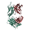
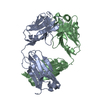
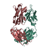
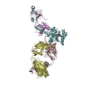
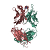
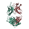
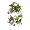
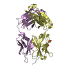
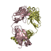
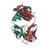
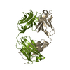
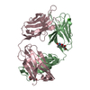
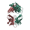
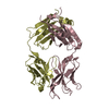
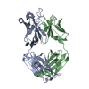
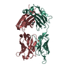
| Entry | Database: PDB / ID: 1flr | ||||||||||||||||||||
|---|---|---|---|---|---|---|---|---|---|---|---|---|---|---|---|---|---|---|---|---|---|
| Title | 4-4-20 FAB FRAGMENT | ||||||||||||||||||||
 Components Components | (4-4-20 (IG* Keywords Keywords IMMUNOGLOBULIN IMMUNOGLOBULINFunction / homology |  Function and homology information Function and homology informationpositive regulation of B cell activation / humoral immune response mediated by circulating immunoglobulin / early endosome to late endosome transport / phagocytosis, recognition / positive regulation of type IIa hypersensitivity / regulation of proteolysis / positive regulation of type I hypersensitivity / antibody-dependent cellular cytotoxicity / Fc-gamma receptor I complex binding / phagocytosis, engulfment ...positive regulation of B cell activation / humoral immune response mediated by circulating immunoglobulin / early endosome to late endosome transport / phagocytosis, recognition / positive regulation of type IIa hypersensitivity / regulation of proteolysis / positive regulation of type I hypersensitivity / antibody-dependent cellular cytotoxicity / Fc-gamma receptor I complex binding / phagocytosis, engulfment / IgG immunoglobulin complex / endosome to lysosome transport / positive regulation of endocytosis / antigen processing and presentation / immunoglobulin mediated immune response / immunoglobulin complex, circulating / immunoglobulin receptor binding / positive regulation of phagocytosis / multivesicular body / complement activation, classical pathway / antigen binding / response to bacterium / positive regulation of immune response / antibacterial humoral response / extracellular space / plasma membrane / cytosolSimilarity search - Function Biological species |  Mus musculus (house mouse) Mus musculus (house mouse)Method | X-RAY DIFFRACTION / Resolution: 1.85 Å  Authors AuthorsWhitlow, M. |  CitationJournal: Protein Eng. / Year: 1995 CitationJournal: Protein Eng. / Year: 1995Title: 1.85 A structure of anti-fluorescein 4-4-20 Fab. Authors: Whitlow, M. / Howard, A.J. / Wood, J.F. / Voss Jr., E.W. / Hardman, K.D. #1: Journal: Proteins / Year: 1989Title: Three-Dimensional Structure of a Fluorescein-Fab Complex Crystallized in 2-Methyl-2,4-Pentanediol Authors: Herron, J.N. / He, X. / Mason, M.L. / Voss Junior, E.W. / Edmundson, A.B. #2: Journal: Proteins / Year: 1988Title: Differences in Crystal Properties and Ligand Affinities of an Antifluorescyl Fab (4-4-20) in Two Solvent Systems Authors: Gibson, A.L. / Herron, J.N. / He, X.-M. / Patrick, V.A. / Mason, M.L. / Lin, J.-N. / Kranz, D.M. / Voss Junior, E.W. / Edmundson, A.B. #3: Journal: J.Biol.Chem. / Year: 1989Title: Comparison of Variable Region Primary Structures within an Anti-Fluorescein Idiotype Family Authors: Bedzyk, W.D. / Johnson, L.S. / Riordon, G.S. / Voss, E.W. History |
|
- Structure visualization
| Structure viewer | Molecule: MolmilJmol/JSmol |
|---|
- Downloads & links
-Download
| PDBx/mmCIF format | 1flr.cif.gz | 107.5 KB | Display | PDBx/mmCIF format |
|---|---|---|---|---|
| PDB format | pdb1flr.ent.gz | 79.8 KB | Display | PDB format |
| PDBx/mmJSON format | 1flr.json.gz | Tree view | PDBx/mmJSON format | |
| Others |  Other downloads Other downloads |
-Validation report
| Arichive directory | https://data.pdbj.org/pub/pdb/validation_reports/fl/1flrftp://data.pdbj.org/pub/pdb/validation_reports/fl/1flr | HTTPS FTP |
|---|
-Related structure data
| Similar structure data |
|---|


-Links
 PDBj
PDBj


- Assembly
| Deposited unit | 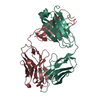
| ||||||||
|---|---|---|---|---|---|---|---|---|---|
| 1 |
| ||||||||
| Unit cell |
| ||||||||
| Atom site foot note | 1: CIS PROLINE - PRO L 8 / 2: CIS PROLINE - PRO L 100 / 3: CIS PROLINE - PRO L 146 / 4: CIS PROLINE - PRO H 152 / 5: CIS PROLINE - PRO H 194 / 6: WATER MOLECULE 870 IS DISORDERED. |
-Components
| #1: Antibody | Mass: 24173.820 Da / Num. of mol.: 1 / Source method: isolated from a natural source / Source: (natural) Mus musculus (house mouse) / Cell: LYMPHOCYTE-PLASMA CELL / Cell line: 4-4-20 MURINE-MURINE HYBRIDOMA / Organ: SPLEEN / Variant: BALB/CV / Strain: BALB/C / References: GenBank: 1589925 |
|---|---|
| #2: Antibody | Mass: 23973.781 Da / Num. of mol.: 1 / Source method: isolated from a natural source / Source: (natural) Mus musculus (house mouse) / Cell: LYMPHOCYTE-PLASMA CELL / Cell line: 4-4-20 MURINE-MURINE HYBRIDOMA / Organ: SPLEEN / Variant: BALB/CV / Strain: BALB/C / References: UniProt: P01865 |
| #3: Chemical | ChemComp-FLU / Fluorescein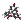  Mass: 332.306 Da / Num. of mol.: 1 / Source method: obtained synthetically / Formula: C20H12O5 Mass: 332.306 Da / Num. of mol.: 1 / Source method: obtained synthetically / Formula: C20H12O5 |
| #4: Water | ChemComp-HOH / Water Mass: 18.015 Da / Num. of mol.: 290 / Source method: isolated from a natural source / Formula: H2O Mass: 18.015 Da / Num. of mol.: 290 / Source method: isolated from a natural source / Formula: H2O |
-Experimental details
-Experiment
| Experiment | Method: X-RAY DIFFRACTION |
|---|
- Sample preparation
| Crystal | Density Matthews: 2.23 Å3/Da / Density % sol: 44.88 % | ||||||||||||||||||||
|---|---|---|---|---|---|---|---|---|---|---|---|---|---|---|---|---|---|---|---|---|---|
| Crystal grow | *PLUS Temperature: 15 ℃ / Method: unknownDetails: Gibson, A.L., (1988) Proteins: Struct.,Funct., Genet., 3, 155. PH range low: 7.3 / PH range high: 6.8 | ||||||||||||||||||||
| Components of the solutions | *PLUS
|
-Data collection
| Diffraction source | Wavelength: 1.54 |
|---|---|
| Detector | Type: SIEMENS-NICOLET X100 / Detector: AREA DETECTOR / Date: Oct 21, 1988 |
| Radiation | Monochromatic (M) / Laue (L): M / Scattering type: x-ray |
| Radiation wavelength | Wavelength: 1.54 Å / Relative weight: 1 |
| Reflection | Num. obs: 29963 / % possible obs: 70 % / Observed criterion σ(I): 0 / Rmerge(I) obs: 0.041 |
| Reflection | *PLUS Highest resolution: 1.79 Å / Num. measured all: 76616 / Rmerge(I) obs: 0.041 |
- Processing
| Software |
| ||||||||||||||||||||||||||||||||||||||||||||||||||||||||||||||||||||||||||||||||||||
|---|---|---|---|---|---|---|---|---|---|---|---|---|---|---|---|---|---|---|---|---|---|---|---|---|---|---|---|---|---|---|---|---|---|---|---|---|---|---|---|---|---|---|---|---|---|---|---|---|---|---|---|---|---|---|---|---|---|---|---|---|---|---|---|---|---|---|---|---|---|---|---|---|---|---|---|---|---|---|---|---|---|---|---|---|---|
| Refinement | Resolution: 1.85→10 Å / σ(F): 2 Details: RESIDUES CYS H 133 TO GLY H 138 ARE IN WEAK ELECTRON DENSITY. THERE IS A DISCONTINUITY IN THE ELECTION DENSITY MAP BETWEEN RESIDUES VAL H 132 AND CYS H 133. THE RESULTING COORDINATES, AS ...Details: RESIDUES CYS H 133 TO GLY H 138 ARE IN WEAK ELECTRON DENSITY. THERE IS A DISCONTINUITY IN THE ELECTION DENSITY MAP BETWEEN RESIDUES VAL H 132 AND CYS H 133. THE RESULTING COORDINATES, AS PRESENTED IN THIS ENTRY, CONTAIN TWO POSITIONS FOR N CYS H 133, ONE OF WHICH IS PROPERLY CONNECTED TO C VAL H 132 BUT QUITE DISTANT (5.58 ANGSTROMS) FROM CA CYS H 133. THE OTHER POSITION FOR N CYS H 133 IS PROPERLY CONNECTED TO CA H 133 BUT QUITE DISTANT (4.51 ANGSTROMS) FROM C VAL H 132.
| ||||||||||||||||||||||||||||||||||||||||||||||||||||||||||||||||||||||||||||||||||||
| Refinement step | Cycle: LAST / Resolution: 1.85→10 Å
| ||||||||||||||||||||||||||||||||||||||||||||||||||||||||||||||||||||||||||||||||||||
| Refine LS restraints |
|
