 Movie
Movie Controller


+ Open data

- Basic information
| Entry | Database: PDB / ID: 1flj | ||||||
|---|---|---|---|---|---|---|---|
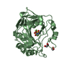
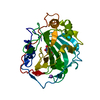
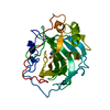
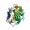
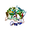
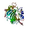
| Title | CRYSTAL STRUCTURE OF S-GLUTATHIOLATED CARBONIC ANHYDRASE III | ||||||
 Components Components | CARBONIC ANHYDRASE III Carbonic anhydrase Carbonic anhydrase | ||||||
 Keywords Keywords | LYASE / Carbonic Anhydrase III / Glutathione / S-Glutathiolated / S-Glutathionylated | ||||||
| Function / homology |  Function and homology information Function and homology informationReversible hydration of carbon dioxide / hydro-lyase activity / phosphatase activity / nickel cation binding / response to bacterium / carbonic anhydrase / carbonate dehydratase activity / one-carbon metabolic process / response to ethanol / response to oxidative stress ...Reversible hydration of carbon dioxide / hydro-lyase activity / phosphatase activity / nickel cation binding / response to bacterium / carbonic anhydrase / carbonate dehydratase activity / one-carbon metabolic process / response to ethanol / response to oxidative stress / zinc ion binding / cytosolSimilarity search - Function | ||||||
| Biological species |  Rattus norvegicus (Norway rat) Rattus norvegicus (Norway rat) | ||||||
| Method | X-RAY DIFFRACTION / Resolution: 1.8 Å | ||||||
 Authors Authors | Mallis, R.J. / Poland, B.W. / Chatterjee, T.K. / Fisher, R.A. / Darmawan, S. / Honzatko, R.B. / Thomas, J.A. | ||||||
 Citation Citation | Journal: FEBS Lett. / Year: 2000 Title: Crystal structure of S-glutathiolated carbonic anhydrase III. Authors: Mallis, R.J. / Poland, B.W. / Chatterjee, T.K. / Fisher, R.A. / Darmawan, S. / Honzatko, R.B. / Thomas, J.A. #1: Journal: Arch.Biochem.Biophys. / Year: 1995Title: Protein Sulfhydryls and Their Role in the Antioxidant Function of Protein S-thiolation Authors: Thomas, J.A. / Poland, B. / Honzatko, R. | ||||||
| History |
|
- Structure visualization
| Structure viewer | Molecule: MolmilJmol/JSmol |
|---|
- Downloads & links
-Download
| PDBx/mmCIF format | 1flj.cif.gz | 75.9 KB | Display | PDBx/mmCIF format |
|---|---|---|---|---|
| PDB format | pdb1flj.ent.gz | 61.2 KB | Display | PDB format |
| PDBx/mmJSON format | 1flj.json.gz | Tree view | PDBx/mmJSON format | |
| Others |  Other downloads Other downloads |
-Validation report
| Arichive directory | https://data.pdbj.org/pub/pdb/validation_reports/fl/1fljftp://data.pdbj.org/pub/pdb/validation_reports/fl/1flj | HTTPS FTP |
|---|
-Related structure data
| Similar structure data |
|---|


-Links
 PDBj
PDBj

- Assembly
| Deposited unit | 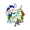
| ||||||||||
|---|---|---|---|---|---|---|---|---|---|---|---|
| 1 |
| ||||||||||
| Unit cell |
|
-Components
| #1: Protein | Carbonic anhydrase / E.C.4.2.1.1 / CARBONATE DEHYDRATASE III / CA-III Mass: 29369.273 Da / Num. of mol.: 1 / Source method: isolated from a natural source / Source: (natural) Rattus norvegicus (Norway rat) / Organ: LIVER / References: UniProt: P14141, carbonic anhydrase | ||
|---|---|---|---|
| #2: Chemical | ChemComp-ZN /   Mass: 65.409 Da / Num. of mol.: 1 / Source method: obtained synthetically / Formula: Zn Mass: 65.409 Da / Num. of mol.: 1 / Source method: obtained synthetically / Formula: Zn | ||
| #3: Chemical | Glutathione  Mass: 307.323 Da / Num. of mol.: 2 / Source method: obtained synthetically / Formula: C10H17N3O6S Mass: 307.323 Da / Num. of mol.: 2 / Source method: obtained synthetically / Formula: C10H17N3O6S#4: Water | ChemComp-HOH / | Water Mass: 18.015 Da / Num. of mol.: 216 / Source method: isolated from a natural source / Formula: H2O Mass: 18.015 Da / Num. of mol.: 216 / Source method: isolated from a natural source / Formula: H2O |
-Experimental details
-Experiment
| Experiment | Method: X-RAY DIFFRACTION / Number of used crystals: 1 |
|---|
- Sample preparation
| Crystal | Density Matthews: 2.07 Å3/Da / Density % sol: 40.58 % | ||||||||||||||||||||||||||||||||||||||||
|---|---|---|---|---|---|---|---|---|---|---|---|---|---|---|---|---|---|---|---|---|---|---|---|---|---|---|---|---|---|---|---|---|---|---|---|---|---|---|---|---|---|
| Crystal grow | Temperature: 298 K / Method: vapor diffusion, hanging drop / pH: 7.5 Details: PEG 3350, propanol, HEPES, pH 7.5, VAPOR DIFFUSION, HANGING DROP, temperature 298K | ||||||||||||||||||||||||||||||||||||||||
| Crystal grow | *PLUS | ||||||||||||||||||||||||||||||||||||||||
| Components of the solutions | *PLUS
|
-Data collection
| Diffraction | Mean temperature: 298 K |
|---|---|
| Diffraction source | Source: ROTATING ANODE / Type: SIEMENS / Wavelength: 1.5418 |
| Detector | Type: SIEMENS / Detector: AREA DETECTOR / Date: May 1, 1994 |
| Radiation | Protocol: SINGLE WAVELENGTH / Monochromatic (M) / Laue (L): M / Scattering type: x-ray |
| Radiation wavelength | Wavelength: 1.5418 Å / Relative weight: 1 |
| Reflection | Resolution: 1.8→5 Å / Num. all: 20193 / Num. obs: 20193 / % possible obs: 90 % / Observed criterion σ(F): 0 / Observed criterion σ(I): 0 / Redundancy: 2.216 % / Biso Wilson estimate: 13 Å2 / Rmerge(I) obs: 0.014 / Net I/σ(I): 5.4 |
| Reflection | *PLUS Lowest resolution: 5 Å / % possible obs: 90 % / Num. measured all: 44741 / Rmerge(I) obs: 0.054 |
- Processing
| Software |
| |||||||||||||||||||||||||
|---|---|---|---|---|---|---|---|---|---|---|---|---|---|---|---|---|---|---|---|---|---|---|---|---|---|---|
| Refinement | Resolution: 1.8→5 Å / Cross valid method: THROUGHOUT / σ(F): 0 / σ(I): 0 / Stereochemistry target values: Engh & Huber
| |||||||||||||||||||||||||
| Refinement step | Cycle: LAST / Resolution: 1.8→5 Å
| |||||||||||||||||||||||||
| Refine LS restraints |
| |||||||||||||||||||||||||
| Software | *PLUS Name: X-PLOR / Classification: refinement | |||||||||||||||||||||||||
| Refinement | *PLUS Num. reflection obs: 14426 / σ(F): 0 / % reflection Rfree: 10 % | |||||||||||||||||||||||||
| Solvent computation | *PLUS | |||||||||||||||||||||||||
| Displacement parameters | *PLUS |
