 Movie
Movie Controller

[English] 日本語
 Yorodumi
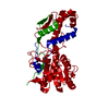
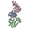
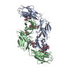
- PDB-1fi4: THE X-RAY CRYSTAL STRUCTURE OF MEVALONATE 5-DIPHOSPHATE DECARBOXY... -
+ Open data

- Basic information
| Entry | Database: PDB / ID: 1fi4 | ||||||
|---|---|---|---|---|---|---|---|
| Title | THE X-RAY CRYSTAL STRUCTURE OF MEVALONATE 5-DIPHOSPHATE DECARBOXYLASE AT 2.3 ANGSTROM RESOLUTION. | ||||||
 Components Components | MEVALONATE 5-DIPHOSPHATE DECARBOXYLASE | ||||||
 Keywords Keywords |  LYASE / mixed alpha/beta structure / ATP binding / decarboxylase / cholesterol biosynthesis / structural genomics / PSI / Protein Structure Initiative / New York SGX Research Center for Structural Genomics / NYSGXRC LYASE / mixed alpha/beta structure / ATP binding / decarboxylase / cholesterol biosynthesis / structural genomics / PSI / Protein Structure Initiative / New York SGX Research Center for Structural Genomics / NYSGXRC | ||||||
| Function / homology |  Function and homology information Function and homology informationSynthesis of Dolichyl-phosphate / Cholesterol biosynthesis / diphosphomevalonate decarboxylase / diphosphomevalonate decarboxylase activity / isopentenyl diphosphate biosynthetic process, mevalonate pathway / sterol biosynthetic process / ATP binding / cytosol / cytoplasmSimilarity search - Function | ||||||
| Biological species |  Saccharomyces cerevisiae (brewer's yeast) Saccharomyces cerevisiae (brewer's yeast) | ||||||
| Method | X-RAY DIFFRACTION / SYNCHROTRON / Resolution: 2.27 Å | ||||||
 Authors Authors | Bonanno, J.B. / Edo, C. / Eswar, N. / Pieper, U. / Romanowski, M.J. / Ilyin, V. / Gerchman, S.E. / Kycia, H. / Studier, F.W. / Sali, A. ...Bonanno, J.B. / Edo, C. / Eswar, N. / Pieper, U. / Romanowski, M.J. / Ilyin, V. / Gerchman, S.E. / Kycia, H. / Studier, F.W. / Sali, A. / Burley, S.K. / New York SGX Research Center for Structural Genomics (NYSGXRC) | ||||||
 Citation Citation | Journal: Proc.Natl.Acad.Sci.USA / Year: 2001 Title: Structural genomics of enzymes involved in sterol/isoprenoid biosynthesis. Authors: Bonanno, J.B. / Edo, C. / Eswar, N. / Pieper, U. / Romanowski, M.J. / Ilyin, V. / Gerchman, S.E. / Kycia, H. / Studier, F.W. / Sali, A. / Burley, S.K. | ||||||
| History |
|
- Structure visualization
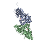
| Structure viewer | Molecule: MolmilJmol/JSmol |
|---|
- Downloads & links
-Download
| PDBx/mmCIF format | 1fi4.cif.gz | 89.7 KB | Display | PDBx/mmCIF format |
|---|---|---|---|---|
| PDB format | pdb1fi4.ent.gz | 71.9 KB | Display | PDB format |
| PDBx/mmJSON format | 1fi4.json.gz | Tree view | PDBx/mmJSON format | |
| Others |  Other downloads Other downloads |
-Validation report
| Arichive directory | https://data.pdbj.org/pub/pdb/validation_reports/fi/1fi4ftp://data.pdbj.org/pub/pdb/validation_reports/fi/1fi4 | HTTPS FTP |
|---|
-Related structure data


-Links
 PDBj
PDBj- Assembly
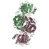
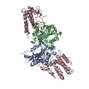
| Deposited unit | 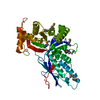
| ||||||||
|---|---|---|---|---|---|---|---|---|---|
| 1 | 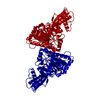
| ||||||||
| Unit cell |
| ||||||||
| Details | physiological dimer observed in crystal |
-Components
| #1: Protein | Mass: 46804.043 Da / Num. of mol.: 1 Mutation: M1(MSE), M89(MSE), M169(MSE), M179(MSE), M192(MSE), M212(MSE), M237(MSE), M254(MSE), M255(MSE), M274(MSE) Source method: isolated from a genetically manipulated source Source: (gene. exp.) Saccharomyces cerevisiae (brewer's yeast)Description: HIS-TAG MODIFIED PET28A PLASMID / Production host:  Escherichia coli (E. coli) Escherichia coli (E. coli)References: UniProt: P32377, diphosphomevalonate decarboxylase |
|---|---|
| #2: Water | ChemComp-HOH / Water Mass: 18.015 Da / Num. of mol.: 164 / Source method: isolated from a natural source / Formula: H2O Mass: 18.015 Da / Num. of mol.: 164 / Source method: isolated from a natural source / Formula: H2O |
-Experimental details
-Experiment
| Experiment | Method: X-RAY DIFFRACTION / Number of used crystals: 1 |
|---|
- Sample preparation
| Crystal | Density Matthews: 2.51 Å3/Da / Density % sol: 51.04 % | ||||||||||||||||||||||||||||||
|---|---|---|---|---|---|---|---|---|---|---|---|---|---|---|---|---|---|---|---|---|---|---|---|---|---|---|---|---|---|---|---|
| Crystal grow | Temperature: 277 K / Method: vapor diffusion, hanging drop / pH: 8.5 Details: 100 mM Tris-HCl pH 8.5, 15% PEG 4K, 1M NaCl, 5% glycerol, 5% ethylene glycol, VAPOR DIFFUSION, HANGING DROP | ||||||||||||||||||||||||||||||
| Crystal grow | *PLUS | ||||||||||||||||||||||||||||||
| Components of the solutions | *PLUS
|
-Data collection
| Diffraction | Mean temperature: 100 K |
|---|---|
| Diffraction source | Source: SYNCHROTRON / Site: NSLS  / Beamline: X25 / Wavelength: 0.979 / Beamline: X25 / Wavelength: 0.979 |
| Detector | Type: BRANDEIS - B4 / Detector: CCD / Date: Oct 13, 1999 |
| Radiation | Protocol: SINGLE WAVELENGTH / Monochromatic (M) / Laue (L): M / Scattering type: x-ray |
| Radiation wavelength | Wavelength: 0.979 Å / Relative weight: 1 |
| Reflection | Resolution: 2.25→30 Å / Num. all: 41954 / Num. obs: 40942 / % possible obs: 97.6 % / Observed criterion σ(F): 9999 / Observed criterion σ(I): 2 / Redundancy: 7.3 % / Biso Wilson estimate: 41 Å2 / Rmerge(I) obs: 0.046 / Net I/σ(I): 25 |
| Reflection shell | Resolution: 2.25→2.33 Å / Redundancy: 6.5 % / Rmerge(I) obs: 0.227 / Num. unique all: 4206 / % possible all: 90.8 |
| Reflection | *PLUS Lowest resolution: 18 Å / % possible obs: 99.1 % / Rmerge(I) obs: 0.03 |
| Reflection shell | *PLUS % possible obs: 90.3 % |
- Processing
| Software |
| |||||||||||||||||||||||||
|---|---|---|---|---|---|---|---|---|---|---|---|---|---|---|---|---|---|---|---|---|---|---|---|---|---|---|
| Refinement | Resolution: 2.27→30 Å / σ(F): 9999 / σ(I): 0 / Stereochemistry target values: Engh & Huber Details: The model was refined against a target of maximum likelihood on solvent flattened phases except for the last round of Powell minimization when a target of maximum likelihood on F was used. ...Details: The model was refined against a target of maximum likelihood on solvent flattened phases except for the last round of Powell minimization when a target of maximum likelihood on F was used. The data were corrected for anomalous scattering by the selenium atoms.
| |||||||||||||||||||||||||
| Refinement step | Cycle: LAST / Resolution: 2.27→30 Å
| |||||||||||||||||||||||||
| Refine LS restraints |
| |||||||||||||||||||||||||
| Software | *PLUS Name: CNS / Version: 0.9 / Classification: refinement | |||||||||||||||||||||||||
| Refinement | *PLUS Lowest resolution: 18 Å / σ(F): 9999 / Rfactor obs: 0.239 | |||||||||||||||||||||||||
| Solvent computation | *PLUS | |||||||||||||||||||||||||
| Displacement parameters | *PLUS | |||||||||||||||||||||||||
| Refine LS restraints | *PLUS Type: c_angle_deg / Dev ideal: 1.3 |
