 Movie
Movie Controller

[English] 日本語
 Yorodumi
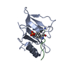
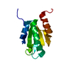
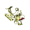
- PDB-1f1w: SRC SH2 THREF1TRP MUTANT COMPLEXED WITH THE PHOSPHOPEPTIDE S(PTR)VNVQN -
+ Open data

- Basic information
| Entry | Database: PDB / ID: 1f1w | ||||||
|---|---|---|---|---|---|---|---|
| Title | SRC SH2 THREF1TRP MUTANT COMPLEXED WITH THE PHOSPHOPEPTIDE S(PTR)VNVQN | ||||||
 Components Components |
| ||||||
 Keywords Keywords |  TRANSFERASE / Src / SH2 domain / phosphopeptide / specificity switch TRANSFERASE / Src / SH2 domain / phosphopeptide / specificity switch | ||||||
| Function / homology |  Function and homology information Function and homology informationSignaling by ERBB2 / Nuclear signaling by ERBB4 / PIP3 activates AKT signaling / Signaling by SCF-KIT / Regulation of KIT signaling / Signaling by EGFR / GAB1 signalosome / Regulation of gap junction activity / FCGR activation / PECAM1 interactions ...Signaling by ERBB2 / Nuclear signaling by ERBB4 / PIP3 activates AKT signaling / Signaling by SCF-KIT / Regulation of KIT signaling / Signaling by EGFR / GAB1 signalosome / Regulation of gap junction activity / FCGR activation / PECAM1 interactions / CD28 co-stimulation / CTLA4 inhibitory signaling / EPHA-mediated growth cone collapse / Ephrin signaling / G alpha (i) signalling events / GP1b-IX-V activation signalling / Recycling pathway of L1 / Thrombin signalling through proteinase activated receptors (PARs) / VEGFR2 mediated cell proliferation / RAF activation / PI5P, PP2A and IER3 Regulate PI3K/AKT Signaling / RET signaling / Receptor Mediated Mitophagy / ADP signalling through P2Y purinoceptor 1 / Downregulation of ERBB4 signaling / EPH-ephrin mediated repulsion of cells / Cyclin D associated events in G1 / Regulation of RUNX3 expression and activity / Activated NTRK3 signals through PI3K / Downstream signal transduction / MAP2K and MAPK activation / Integrin signaling / GRB2:SOS provides linkage to MAPK signaling for Integrins / MET activates PTK2 signaling / Extra-nuclear estrogen signaling / EPHB-mediated forward signaling / p130Cas linkage to MAPK signaling for integrins / VEGFA-VEGFR2 Pathway / connexin binding / osteoclast development / progesterone receptor signaling pathway / negative regulation of intrinsic apoptotic signaling pathway / bone resorption / extrinsic component of cytoplasmic side of plasma membrane / negative regulation of extrinsic apoptotic signaling pathway / non-specific protein-tyrosine kinase / non-membrane spanning protein tyrosine kinase activity / epidermal growth factor receptor signaling pathway / cell junction / protein phosphatase binding / protein tyrosine kinase activity / mitochondrial inner membrane / cell differentiation / cytoskeleton / endosome membrane / regulation of cell cycle / cell adhesion / cell cycle / phosphorylation / signaling receptor binding / focal adhesion / innate immune response / heme binding / perinuclear region of cytoplasm / protein-containing complex / ATP binding / membrane / nucleus / plasma membrane / cytosolSimilarity search - Function | ||||||
| Biological species |  Gallus gallus (chicken) Gallus gallus (chicken) | ||||||
| Method | X-RAY DIFFRACTION / Resolution: 2.1 Å | ||||||
 Authors Authors | Kimber, M.S. / Nachman, J. / Cunningham, A.M. / Gish, G.D. / Pawson, T. / Pai, E.F. | ||||||
 Citation Citation | Journal: Mol.Cell / Year: 2000 Title: Structural basis for specificity switching of the Src SH2 domain. Authors: Kimber, M.S. / Nachman, J. / Cunningham, A.M. / Gish, G.D. / Pawson, T. / Pai, E.F. | ||||||
| History |
|
- Structure visualization
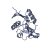
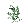
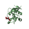
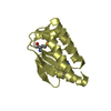
| Structure viewer | Molecule: MolmilJmol/JSmol |
|---|
- Downloads & links
-Download
| PDBx/mmCIF format | 1f1w.cif.gz | 37.7 KB | Display | PDBx/mmCIF format |
|---|---|---|---|---|
| PDB format | pdb1f1w.ent.gz | 25 KB | Display | PDB format |
| PDBx/mmJSON format | 1f1w.json.gz | Tree view | PDBx/mmJSON format | |
| Others |  Other downloads Other downloads |
-Validation report
| Arichive directory | https://data.pdbj.org/pub/pdb/validation_reports/f1/1f1wftp://data.pdbj.org/pub/pdb/validation_reports/f1/1f1w | HTTPS FTP |
|---|
-Related structure data
| Related structure data | 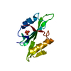 1f2fC 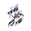 1spsS C: citing same article ( S: Starting model for refinement |
|---|---|
| Similar structure data |


-Links
 PDBj
PDBj- Assembly
| Deposited unit | 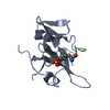
| ||||||||
|---|---|---|---|---|---|---|---|---|---|
| 1 |
| ||||||||
| 2 | 
| ||||||||
| 3 | 
| ||||||||
| Unit cell |
|
-Components
| #1: Protein | Mass: 12058.653 Da / Num. of mol.: 1 / Fragment: SRC SH2 DOMAIN / Mutation: T215W Source method: isolated from a genetically manipulated source Source: (gene. exp.) Gallus gallus (chicken) / Plasmid: PET11D / Production host:  Escherichia coli (E. coli) / Strain (production host): BL21(DE3)PLYSS / References: UniProt: P00523, EC: 2.7.1.112 Escherichia coli (E. coli) / Strain (production host): BL21(DE3)PLYSS / References: UniProt: P00523, EC: 2.7.1.112 |
|---|---|
| #2: Protein/peptide | Mass: 902.842 Da / Num. of mol.: 1 / Source method: obtained synthetically / Details: chemically synthesized |
| #3: Water | ChemComp-HOH / Water Mass: 18.015 Da / Num. of mol.: 75 / Source method: isolated from a natural source / Formula: H2O Mass: 18.015 Da / Num. of mol.: 75 / Source method: isolated from a natural source / Formula: H2O |
-Experimental details
-Experiment
| Experiment | Method: X-RAY DIFFRACTION / Number of used crystals: 1 |
|---|
- Sample preparation
| Crystal | Density Matthews: 1.9 Å3/Da / Density % sol: 35.19 % | |||||||||||||||
|---|---|---|---|---|---|---|---|---|---|---|---|---|---|---|---|---|
| Crystal grow | Temperature: 298 K / Method: vapor diffusion, hanging drop / pH: 5.6 Details: 1.2 M Na Citrate, pH 5.6, VAPOR DIFFUSION, HANGING DROP, temperature 298.0K | |||||||||||||||
| Crystal grow | *PLUS Details: 1:1.5 molar ratio of protein/peptide complex | |||||||||||||||
| Components of the solutions | *PLUS
|
-Data collection
| Diffraction | Mean temperature: 100 K |
|---|---|
| Diffraction source | Source: ROTATING ANODE / Type: RIGAKU FR-C / Wavelength: 1.5418 |
| Detector | Type: MARRESEARCH / Detector: IMAGE PLATE / Date: Jan 6, 1999 |
| Radiation | Protocol: SINGLE WAVELENGTH / Monochromatic (M) / Laue (L): M / Scattering type: x-ray |
| Radiation wavelength | Wavelength: 1.5418 Å / Relative weight: 1 |
| Reflection | Resolution: 2.1→12 Å / Num. all: 14854 / Num. obs: 5813 / % possible obs: 95 % / Observed criterion σ(I): -3 / Redundancy: 3.2 % / Biso Wilson estimate: 13.3 Å2 / Rmerge(I) obs: 0.092 / Net I/σ(I): 9.5 |
- Processing
| Software |
| ||||||||||||||||||||||||||||||||||||
|---|---|---|---|---|---|---|---|---|---|---|---|---|---|---|---|---|---|---|---|---|---|---|---|---|---|---|---|---|---|---|---|---|---|---|---|---|---|
| Refinement | Starting model: 1sps Resolution: 2.1→12 Å / Rfactor Rfree error: 0.011 / Data cutoff high absF: 218589.02 / Data cutoff low absF: 0 / Isotropic thermal model: RESTRAINED / Cross valid method: THROUGHOUT / σ(F): 0 / Stereochemistry target values: Engh & Huber
| ||||||||||||||||||||||||||||||||||||
| Solvent computation | Solvent model: FLAT MODEL / Bsol: 31.73 Å2 / ksol: 0.3842 e/Å3 | ||||||||||||||||||||||||||||||||||||
| Displacement parameters | Biso mean: 20.5 Å2
| ||||||||||||||||||||||||||||||||||||
| Refine analyze |
| ||||||||||||||||||||||||||||||||||||
| Refinement step | Cycle: LAST / Resolution: 2.1→12 Å
| ||||||||||||||||||||||||||||||||||||
| Refine LS restraints |
| ||||||||||||||||||||||||||||||||||||
| LS refinement shell | Resolution: 2.1→2.23 Å / Rfactor Rfree error: 0.033 / Total num. of bins used: 6
| ||||||||||||||||||||||||||||||||||||
| Xplor file |
| ||||||||||||||||||||||||||||||||||||
| Software | *PLUS Name: CNS / Version: 0.5 / Classification: refinement | ||||||||||||||||||||||||||||||||||||
| Refine LS restraints | *PLUS
|
