Gluconeogenesis / sucrose biosynthetic process / fructose-bisphosphatase / fructose 1,6-bisphosphate 1-phosphatase activity / negative regulation of Ras protein signal transduction / fructose 1,6-bisphosphate metabolic process / cellular response to magnesium ion / fructose 6-phosphate metabolic process / fructose metabolic process / monosaccharide binding ...Gluconeogenesis / sucrose biosynthetic process / fructose-bisphosphatase / fructose 1,6-bisphosphate 1-phosphatase activity / negative regulation of Ras protein signal transduction / fructose 1,6-bisphosphate metabolic process / cellular response to magnesium ion / fructose 6-phosphate metabolic process / fructose metabolic process / monosaccharide binding / negative regulation of glycolytic process / regulation of gluconeogenesis / AMP binding / dephosphorylation / gluconeogenesis / negative regulation of cell growth / cellular response to xenobiotic stimulus / RNA polymerase II-specific DNA-binding transcription factor binding / negative regulation of transcription by RNA polymerase II / identical protein binding / metal ion binding / nucleus / cytosol / cytoplasm Similarity search - Function
Fructose-1,6-bisphosphatase / Fructose-1,6-bisphosphatase, active site / Fructose-1-6-bisphosphatase class 1, C-terminal / Fructose-1-6-bisphosphatase active site. / Fructose-1,6-bisphosphatase class 1 / Fructose-1-6-bisphosphatase class I, N-terminal / Fructose-1-6-bisphosphatase, N-terminal domain / Fructose-1-6-bisphosphatase, C-terminal domain / D-Maltodextrin-Binding Protein; domain 2 - #80 / Fructose-1,6-Bisphosphatase, subunit A, domain 1 ...Fructose-1,6-bisphosphatase / Fructose-1,6-bisphosphatase, active site / Fructose-1-6-bisphosphatase class 1, C-terminal / Fructose-1-6-bisphosphatase active site. / Fructose-1,6-bisphosphatase class 1 / Fructose-1-6-bisphosphatase class I, N-terminal / Fructose-1-6-bisphosphatase, N-terminal domain / Fructose-1-6-bisphosphatase, C-terminal domain / D-Maltodextrin-Binding Protein; domain 2 - #80 / Fructose-1,6-Bisphosphatase, subunit A, domain 1 / Fructose-1,6-Bisphosphatase; Chain A, domain 1 / D-Maltodextrin-Binding Protein; domain 2 / 2-Layer Sandwich / 3-Layer(aba) Sandwich / Alpha Beta Similarity search - Domain/homology
In the structure databanks used in Yorodumi, some data are registered as the other names, "COVID-19 virus" and "2019-nCoV". Here are the details of the virus and the list of structure data.
Jan 31, 2019. EMDB accession codes are about to change! (news from PDBe EMDB page)
EMDB accession codes are about to change! (news from PDBe EMDB page)
The allocation of 4 digits for EMDB accession codes will soon come to an end. Whilst these codes will remain in use, new EMDB accession codes will include an additional digit and will expand incrementally as the available range of codes is exhausted. The current 4-digit format prefixed with “EMD-” (i.e. EMD-XXXX) will advance to a 5-digit format (i.e. EMD-XXXXX), and so on. It is currently estimated that the 4-digit codes will be depleted around Spring 2019, at which point the 5-digit format will come into force.
The EM Navigator/Yorodumi systems omit the EMD- prefix.
Related info.:Q: What is EMD? / ID/Accession-code notation in Yorodumi/EM Navigator
Yorodumi is a browser for structure data from EMDB, PDB, SASBDB, etc.
This page is also the successor to EM Navigator detail page, and also detail information page/front-end page for Omokage search.
The word "yorodu" (or yorozu) is an old Japanese word meaning "ten thousand". "mi" (miru) is to see.
Related info.:EMDB / PDB / SASBDB / Comparison of 3 databanks / Yorodumi Search / Aug 31, 2016. New EM Navigator & Yorodumi / Yorodumi Papers / Jmol/JSmol / Function and homology information / Changes in new EM Navigator and Yorodumi
 Movie
Movie Controller

 Yorodumi
 Open data
 Basic information
 Components
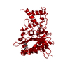
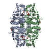
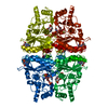
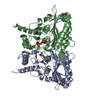
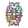
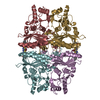
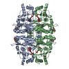
 Fructose 1,6-bisphosphatase
  Keywords
 Function and homology information
 Authors
 Citation
 Structure visualization
 Downloads & links
 Other downloads


 PDBj
PDBj


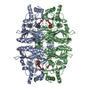
 Assembly


 Type: D-saccharide, beta linking / Mass: 260.136 Da / Num. of mol.: 2


 Mass: 65.409 Da / Num. of mol.: 2 / Source method: obtained synthetically / Formula: Zn
 Mass: 94.971 Da / Num. of mol.: 2 / Source method: obtained synthetically / Formula: PO4
 Mass: 347.221 Da / Num. of mol.: 2 / Source method: obtained synthetically / Formula: C10H14N5O7P / Comment: AMP*YM
 Sample preparation
 Processing