 Movie
Movie Controller


+ Open data

- Basic information
| Entry | Database: PDB / ID: 1ev9 | ||||||
|---|---|---|---|---|---|---|---|
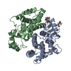
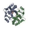
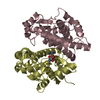
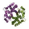
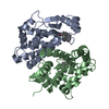
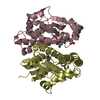
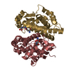
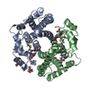
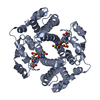
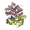
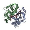
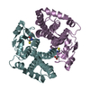
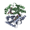
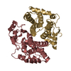
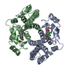
| Title | RAT GLUTATHIONE S-TRANSFERASE A1-1 MUTANT W21F WITH GSO3 BOUND | ||||||
 Components Components | GLUTATHIONE S-TRANSFERASE A1-1 | ||||||
 Keywords Keywords | TRANSFERASE / disordered C-terminal helices | ||||||
| Function / homology |  Function and homology information Function and homology informationAzathioprine ADME / Glutathione conjugation / Heme degradation / Isomerases; Intramolecular oxidoreductases; Transposing C=C bonds / dinitrosyl-iron complex binding / glutathione binding / glutathione derivative biosynthetic process / steroid delta-isomerase activity / linoleic acid metabolic process / glutathione peroxidase activity ...Azathioprine ADME / Glutathione conjugation / Heme degradation / Isomerases; Intramolecular oxidoreductases; Transposing C=C bonds / dinitrosyl-iron complex binding / glutathione binding / glutathione derivative biosynthetic process / steroid delta-isomerase activity / linoleic acid metabolic process / glutathione peroxidase activity / prostaglandin metabolic process / glutathione transferase / glutathione transferase activity / Oxidoreductases; Acting on a peroxide as acceptor; Peroxidases / xenobiotic catabolic process / glutathione metabolic process / epithelial cell differentiation / xenobiotic metabolic process / response to nutrient levels / fatty acid binding / response to xenobiotic stimulus / protein homodimerization activity / cytosolSimilarity search - Function | ||||||
| Biological species |  Rattus norvegicus (Norway rat) Rattus norvegicus (Norway rat) | ||||||
| Method | X-RAY DIFFRACTION / Resolution: 2.2 Å | ||||||
 Authors Authors | Adman, E.T. / Le Trong, I. / Stenkamp, R.E. / Nieslanik, B.S. / Dietze, E.C. / Tai, G. / Ibarra, C. / Atkins, W.M. | ||||||
 Citation Citation | Journal: Proteins / Year: 2001 Title: Localization of the C-terminus of rat glutathione S-transferase A1-1: crystal structure of mutants W21F and W21F/F220Y. Authors: Adman, E.T. / Le Trong, I. / Stenkamp, R.E. / Nieslanik, B.S. / Dietze, E.C. / Tai, G. / Ibarra, C. / Atkins, W.M. | ||||||
| History |
|
- Structure visualization
| Structure viewer | Molecule: MolmilJmol/JSmol |
|---|
- Downloads & links
-Download
| PDBx/mmCIF format | 1ev9.cif.gz | 142.8 KB | Display | PDBx/mmCIF format |
|---|---|---|---|---|
| PDB format | pdb1ev9.ent.gz | 113.9 KB | Display | PDB format |
| PDBx/mmJSON format | 1ev9.json.gz | Tree view | PDBx/mmJSON format | |
| Others |  Other downloads Other downloads |
-Validation report
| Arichive directory | https://data.pdbj.org/pub/pdb/validation_reports/ev/1ev9ftp://data.pdbj.org/pub/pdb/validation_reports/ev/1ev9 | HTTPS FTP |
|---|
-Related structure data


-Links
 PDBj
PDBj

- Assembly
| Deposited unit | 
| ||||||||
|---|---|---|---|---|---|---|---|---|---|
| 1 | 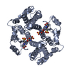
| ||||||||
| 2 | 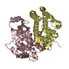
| ||||||||
| Unit cell |
|
-Components
| #1: Protein | Mass: 25464.885 Da / Num. of mol.: 3 / Mutation: W20F Source method: isolated from a genetically manipulated source Source: (gene. exp.) Rattus norvegicus (Norway rat) / Plasmid: PKKGTB34-W21F / Production host:  Escherichia coli (E. coli) / References: UniProt: P00502, glutathione transferase Escherichia coli (E. coli) / References: UniProt: P00502, glutathione transferase#2: Chemical | Sulfate  Mass: 96.063 Da / Num. of mol.: 3 / Source method: obtained synthetically / Formula: SO4 Mass: 96.063 Da / Num. of mol.: 3 / Source method: obtained synthetically / Formula: SO4#3: Chemical |   Mass: 355.322 Da / Num. of mol.: 3 / Source method: obtained synthetically / Formula: C10H17N3O9S Mass: 355.322 Da / Num. of mol.: 3 / Source method: obtained synthetically / Formula: C10H17N3O9S#4: Water | ChemComp-HOH / | Water Mass: 18.015 Da / Num. of mol.: 166 / Source method: isolated from a natural source / Formula: H2O Mass: 18.015 Da / Num. of mol.: 166 / Source method: isolated from a natural source / Formula: H2O |
|---|
-Experimental details
-Experiment
| Experiment | Method: X-RAY DIFFRACTION / Number of used crystals: 1 |
|---|
- Sample preparation
| Crystal | Density Matthews: 2.69 Å3/Da / Density % sol: 54.22 % | ||||||||||||||||||||||||||||||
|---|---|---|---|---|---|---|---|---|---|---|---|---|---|---|---|---|---|---|---|---|---|---|---|---|---|---|---|---|---|---|---|
| Crystal grow | Temperature: 298 K / Method: vapor diffusion, sitting drop / pH: 10.5 Details: 0.2M lithium sulfate, 50% saturated ammonium sulfate, 0.1M 3-cyclohexylamino-1-propane sulfonic acid (CAPS), pH 10.5, VAPOR DIFFUSION, SITTING DROP, temperature 298K | ||||||||||||||||||||||||||||||
| Crystal grow | *PLUS | ||||||||||||||||||||||||||||||
| Components of the solutions | *PLUS
|
-Data collection
| Diffraction | Mean temperature: 110 K |
|---|---|
| Diffraction source | Source: ROTATING ANODE / Type: RIGAKU RU200 / Wavelength: 1.5418 |
| Detector | Type: RIGAKU RAXIS IIC / Detector: IMAGE PLATE / Date: Sep 29, 1997 |
| Radiation | Protocol: SINGLE WAVELENGTH / Monochromatic (M) / Laue (L): M / Scattering type: x-ray |
| Radiation wavelength | Wavelength: 1.5418 Å / Relative weight: 1 |
| Reflection | Resolution: 1.8→20 Å / Num. all: 65590 / Num. obs: 65590 / % possible obs: 82.7 % / Observed criterion σ(F): 0 / Observed criterion σ(I): 0 / Rmerge(I) obs: 0.083 / Net I/σ(I): 13.6 |
| Reflection shell | Resolution: 1.8→1.88 Å / Rmerge(I) obs: 0.926 / Num. unique all: 1822 / % possible all: 18.6 |
| Reflection | *PLUS Highest resolution: 1.98 Å / Num. obs: 57919 / % possible obs: 97 % / Rmerge(I) obs: 0.085 |
| Reflection shell | *PLUS Highest resolution: 1.98 Å / Lowest resolution: 2.1 Å / % possible obs: 88 % / Num. unique obs: 8678 / Rmerge(I) obs: 0.67 / Mean I/σ(I) obs: 1.5 |
- Processing
| Software |
| ||||||||||||||||||||
|---|---|---|---|---|---|---|---|---|---|---|---|---|---|---|---|---|---|---|---|---|---|
| Refinement | Resolution: 2.2→20 Å / σ(F): 5 / σ(I): 10 / Stereochemistry target values: Engh & Huber Details: refined with Xplor 3.8 bulk solvent correction included KSOL = 0.8, BSOL = 20. anisotropic overall B scaling -5.3,9.3,-4.0
| ||||||||||||||||||||
| Refinement step | Cycle: LAST / Resolution: 2.2→20 Å
| ||||||||||||||||||||
| Refine LS restraints |
| ||||||||||||||||||||
| Software | *PLUS Name: X-PLOR / Version: 3.843 / Classification: refinement | ||||||||||||||||||||
| Refinement | *PLUS Highest resolution: 2.2 Å / Lowest resolution: 19.8 Å / σ(F): 5 / % reflection Rfree: 10 % / Rfactor obs: 0.236 | ||||||||||||||||||||
| Solvent computation | *PLUS | ||||||||||||||||||||
| Displacement parameters | *PLUS | ||||||||||||||||||||
| Refine LS restraints | *PLUS Type: x_angle_deg / Dev ideal: 2.5 |
