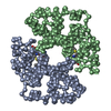
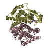
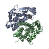
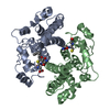
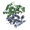
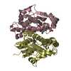
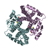
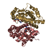
GLUTATHIONE-S-TRANSFERASEA2-2 / GLUTATHIONE-S-TRANSFERASE A2-2 / GTH2 / HA SUBUNIT 2 / GST-GAMMA / GSTA2-2 / GST CLASS-ALPHA MEMBER 2
Mass: 25715.006 Da / Num. of mol.: 8 Source method: isolated from a genetically manipulated source Source: (gene. exp.) HOMO SAPIENS (human) / Plasmid: PET / Production host: ESCHERICHIA COLI (E. coli) / Strain (production host): XL BLUE / References: UniProt: P09210, glutathione transferase
Mass: 18.015 Da / Num. of mol.: 557 / Source method: isolated from a natural source / Formula: H2O
Sequence details
S112T IS A NATURAL VARIANT
-
Experimental details
-
Experiment
Experiment
Method: X-RAY DIFFRACTION / Number of used crystals: 1
-
Sample preparation
Crystal
Density Matthews: 2.39 Å3/Da / Density % sol: 48.06 % / Description: NONE
Crystal grow
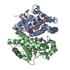
Method: vapor diffusion, hanging drop / pH: 8 Details: THE CRYSTALS WERE OBTAINED BY HANGING DROP VAPOUR TECHNIQUE BY MIXING 5 UL OF RESERVOIR SOLUTION [100 MM TRIS-HCL PH 7.8, 18% (V/V) PEG 4000, AND 2 MM DITHIOTHREITOL] WITH 5 UL OF PROTEIN ...Details: THE CRYSTALS WERE OBTAINED BY HANGING DROP VAPOUR TECHNIQUE BY MIXING 5 UL OF RESERVOIR SOLUTION [100 MM TRIS-HCL PH 7.8, 18% (V/V) PEG 4000, AND 2 MM DITHIOTHREITOL] WITH 5 UL OF PROTEIN SOLUTION (10 MG/ML IN 10 MM TRIS-HCL PH 7.8), 1UL OF 200 MM SPERMINE AND 1 UL OF 25 MM GLUTATHIONE
Resolution: 2.3→42 Å / Cor.coef. Fo:Fc: 0.911 / Cor.coef. Fo:Fc free: 0.857 / SU B: 10.37 / SU ML: 0.255 / Cross valid method: THROUGHOUT / ESU R: 0.588 / ESU R Free: 0.32 / Stereochemistry target values: MAXIMUM LIKELIHOOD / Details: HYDROGENS HAVE BEEN ADDED IN THE RIDING POSITIONS.
Rfactor
Num. reflection
% reflection
Selection details
Rfree
0.29735
4027
5 %
RANDOM
Rwork
0.24558
-
-
-
obs
0.24811
75947
91.97 %
-
Solvent computation
Ion probe radii: 0.8 Å / Shrinkage radii: 0.8 Å / VDW probe radii: 1.2 Å / Solvent model: MASK
 Movie
Movie Controller


 Open data
 Basic information
 Components
 Keywords
 TRANSFERASE /
  Function and homology information
 Authors
 Citation
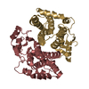
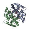
 Structure visualization
 Downloads & links
 Other downloads

 PDBj
PDBj


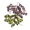
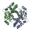
 Assembly


 Mass: 307.323 Da / Num. of mol.: 8 / Source method: obtained synthetically / Formula: C10H17N3O6S
 Mass: 18.015 Da / Num. of mol.: 557 / Source method: isolated from a natural source / Formula: H2O
 Sample preparation
 / Beamline: ID14-1 / Wavelength: 0.933
  Processing