 Movie
Movie Controller

[English] 日本語
 Yorodumi
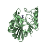
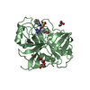
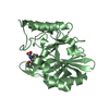
- PDB-1ekb: THE SERINE PROTEASE DOMAIN OF ENTEROPEPTIDASE BOUND TO INHIBITOR ... -
+ Open data

- Basic information
| Entry | Database: PDB / ID: 1ekb | ||||||
|---|---|---|---|---|---|---|---|
| Title | THE SERINE PROTEASE DOMAIN OF ENTEROPEPTIDASE BOUND TO INHIBITOR VAL-ASP-ASP-ASP-ASP-LYS-CHLOROMETHANE | ||||||
 Components Components |
| ||||||
 Keywords Keywords | HYDROLASE/HYDROLASE INHIBITOR /  ENTEROPEPTIDASE / TRYPSINOGEN ACTIVATION / HYDROLASE-HYDROLASE INHIBITOR COMPLEX ENTEROPEPTIDASE / TRYPSINOGEN ACTIVATION / HYDROLASE-HYDROLASE INHIBITOR COMPLEX | ||||||
| Function / homology |  Function and homology information Function and homology informationprotein digestion / enteropeptidase / trypsinogen activation / scavenger receptor activity / membrane => GO:0016020 / serine-type endopeptidase activity / membraneSimilarity search - Function | ||||||
| Biological species |  Bos taurus (cattle) Bos taurus (cattle) | ||||||
| Method | X-RAY DIFFRACTION / MOLECULAR REPLACEMENT / Resolution: 2.3 Å | ||||||
 Authors Authors | Fuetterer, K. / Lu, D. / Sadler, J.E. / Waksman, G. | ||||||
 Citation Citation | Journal: J.Mol.Biol. / Year: 1999 Title: Crystal structure of enteropeptidase light chain complexed with an analog of the trypsinogen activation peptide. Authors: Lu, D. / Futterer, K. / Korolev, S. / Zheng, X. / Tan, K. / Waksman, G. / Sadler, J.E. | ||||||
| History |
|
- Structure visualization
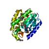
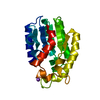
| Structure viewer | Molecule: MolmilJmol/JSmol |
|---|
- Downloads & links
-Download
| PDBx/mmCIF format | 1ekb.cif.gz | 60.6 KB | Display | PDBx/mmCIF format |
|---|---|---|---|---|
| PDB format | pdb1ekb.ent.gz | 46.4 KB | Display | PDB format |
| PDBx/mmJSON format | 1ekb.json.gz | Tree view | PDBx/mmJSON format | |
| Others |  Other downloads Other downloads |
-Validation report
| Arichive directory | https://data.pdbj.org/pub/pdb/validation_reports/ek/1ekbftp://data.pdbj.org/pub/pdb/validation_reports/ek/1ekb | HTTPS FTP |
|---|
-Related structure data
| Related structure data |  1gcdS S: Starting model for refinement |
|---|---|
| Similar structure data |


-Links
 PDBj
PDBj
- Assembly
| Deposited unit | 
| ||||||||
|---|---|---|---|---|---|---|---|---|---|
| 1 |
| ||||||||
| Unit cell |
|
-Components
| #1: Protein/peptide | / E.C.3.4.21.9 / ENTEROKINASE / HEAVY CHAIN Mass: 1419.708 Da / Num. of mol.: 1 Fragment: 13-AMINO ACID REMNANT OF AMINO TERMINAL DOMAIN OF HEAVY CHAIN Source method: isolated from a genetically manipulated source Source: (gene. exp.) Bos taurus (cattle) / Plasmid: PETL / Species (production host): Escherichia coli / Production host:  Escherichia coli BL21(DE3) (bacteria) / Strain (production host): BL21(DE3) / References: UniProt: P98072, enteropeptidase Escherichia coli BL21(DE3) (bacteria) / Strain (production host): BL21(DE3) / References: UniProt: P98072, enteropeptidase | ||||
|---|---|---|---|---|---|
| #2: Protein | / E.C.3.4.21.9 / ENTEROKINASE / LIGHT CHAIN Mass: 26283.785 Da / Num. of mol.: 1 / Fragment: SERINE PROTEASE DOMAIN OR LIGHT CHAIN Source method: isolated from a genetically manipulated source Source: (gene. exp.) Bos taurus (cattle) / Plasmid: PETL / Species (production host): Escherichia coli / Production host: Escherichia coli BL21(DE3) (bacteria) / Strain (production host): BL21(DE3) / References: UniProt: P98072, enteropeptidase | ||||
| #3: Protein/peptide | Mass: 740.158 Da / Num. of mol.: 1 / Source method: obtained synthetically | ||||
| #4: Chemical |   Mass: 65.409 Da / Num. of mol.: 2 / Source method: obtained synthetically / Formula: Zn Mass: 65.409 Da / Num. of mol.: 2 / Source method: obtained synthetically / Formula: Zn#5: Water | ChemComp-HOH / | Water Mass: 18.015 Da / Num. of mol.: 108 / Source method: isolated from a natural source / Formula: H2O Mass: 18.015 Da / Num. of mol.: 108 / Source method: isolated from a natural source / Formula: H2OCompound details | VAL-ASP-ASP-ASP-ASP-LYK-CHLOROMETHANE (CHAIN C) HAS FORMED CONNECTIONS TO ENTEROPETPIDASE: 1) VIA A ...VAL-ASP-ASP-ASP-ASP-LYK-CHLOROMETH | |
-Experimental details
-Experiment
| Experiment | Method: X-RAY DIFFRACTION / Number of used crystals: 1 |
|---|
- Sample preparation
| Crystal | Density Matthews: 2.12 Å3/Da / Density % sol: 40 % | ||||||||||||||||||||||||||||||
|---|---|---|---|---|---|---|---|---|---|---|---|---|---|---|---|---|---|---|---|---|---|---|---|---|---|---|---|---|---|---|---|
| Crystal grow | pH: 7.5 / Details: pH 7.5 | ||||||||||||||||||||||||||||||
| Crystal | *PLUS | ||||||||||||||||||||||||||||||
| Crystal grow | *PLUS Temperature: 20 ℃ / Method: vapor diffusion, hanging drop / pH: 5 | ||||||||||||||||||||||||||||||
| Components of the solutions | *PLUS
|
-Data collection
| Diffraction | Mean temperature: 100 K |
|---|---|
| Diffraction source | Source: ROTATING ANODE / Type: RIGAKU RU200 / Wavelength: 1.5418 |
| Detector | Type: RIGAKU / Detector: IMAGE PLATE / Date: Apr 15, 1998 |
| Radiation | Monochromator: GRAPHITE / Protocol: SINGLE WAVELENGTH / Monochromatic (M) / Laue (L): M / Scattering type: x-ray |
| Radiation wavelength | Wavelength: 1.5418 Å / Relative weight: 1 |
| Reflection | Resolution: 2.3→30 Å / Num. obs: 10541 / % possible obs: 92.6 % / Observed criterion σ(I): 1 / Redundancy: 3 % / Biso Wilson estimate: 32.2 Å2 / Rmerge(I) obs: 0.044 / Rsym value: 0.044 / Net I/σ(I): 10 |
| Reflection shell | Resolution: 2.3→2.38 Å / Redundancy: 2 % / Rmerge(I) obs: 0.088 / Mean I/σ(I) obs: 3 / Rsym value: 0.088 / % possible all: 89.2 |
| Reflection | *PLUS Num. measured all: 28051 |
| Reflection shell | *PLUS % possible obs: 89.2 % |
- Processing
| Software |
| ||||||||||||||||||||||||||||||||||||||||||||||||||||||||||||||||||||||||||||||||
|---|---|---|---|---|---|---|---|---|---|---|---|---|---|---|---|---|---|---|---|---|---|---|---|---|---|---|---|---|---|---|---|---|---|---|---|---|---|---|---|---|---|---|---|---|---|---|---|---|---|---|---|---|---|---|---|---|---|---|---|---|---|---|---|---|---|---|---|---|---|---|---|---|---|---|---|---|---|---|---|---|---|
| Refinement | Method to determine structure: MOLECULAR REPLACEMENT Starting model: PDB ENTRY 1GCD Resolution: 2.3→30 Å / Rfactor Rfree error: 0.012 / Data cutoff high rms absF: 354608.98 / Isotropic thermal model: RESTRAINED / Cross valid method: THROUGHOUT / σ(F): 2
| ||||||||||||||||||||||||||||||||||||||||||||||||||||||||||||||||||||||||||||||||
| Solvent computation | Solvent model: FLAT MODEL / Bsol: 24.18 Å2 / ksol: 0.347 e/Å3 | ||||||||||||||||||||||||||||||||||||||||||||||||||||||||||||||||||||||||||||||||
| Displacement parameters | Biso mean: 23.8 Å2 | ||||||||||||||||||||||||||||||||||||||||||||||||||||||||||||||||||||||||||||||||
| Refine analyze |
| ||||||||||||||||||||||||||||||||||||||||||||||||||||||||||||||||||||||||||||||||
| Refinement step | Cycle: LAST / Resolution: 2.3→30 Å
| ||||||||||||||||||||||||||||||||||||||||||||||||||||||||||||||||||||||||||||||||
| Refine LS restraints |
| ||||||||||||||||||||||||||||||||||||||||||||||||||||||||||||||||||||||||||||||||
| LS refinement shell | Resolution: 2.3→2.4 Å / Rfactor Rfree error: 0.044 / Total num. of bins used: 8
| ||||||||||||||||||||||||||||||||||||||||||||||||||||||||||||||||||||||||||||||||
| Xplor file |
| ||||||||||||||||||||||||||||||||||||||||||||||||||||||||||||||||||||||||||||||||
| Software | *PLUS Name: CNS / Version: 0.5 / Classification: refinement | ||||||||||||||||||||||||||||||||||||||||||||||||||||||||||||||||||||||||||||||||
| Refinement | *PLUS σ(F): 2 / % reflection Rfree: 4.9 % / Rfactor obs: 0.234 | ||||||||||||||||||||||||||||||||||||||||||||||||||||||||||||||||||||||||||||||||
| Solvent computation | *PLUS | ||||||||||||||||||||||||||||||||||||||||||||||||||||||||||||||||||||||||||||||||
| Displacement parameters | *PLUS Biso mean: 23.8 Å2 | ||||||||||||||||||||||||||||||||||||||||||||||||||||||||||||||||||||||||||||||||
| Refine LS restraints | *PLUS
| ||||||||||||||||||||||||||||||||||||||||||||||||||||||||||||||||||||||||||||||||
| LS refinement shell | *PLUS Rfactor Rfree: 0.367 / % reflection Rfree: 5.8 % / Rfactor Rwork: 0.295 |
