 Movie
Movie Controller

[English] 日本語
 Yorodumi
- PDB-1ejj: CRYSTAL STRUCTURAL ANALYSIS OF PHOSPHOGLYCERATE MUTASE COCRYSTALL... -
+ Open data

- Basic information
| Entry | Database: PDB / ID: 1ejj | ||||||
|---|---|---|---|---|---|---|---|
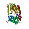
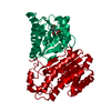
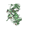
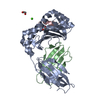
| Title | CRYSTAL STRUCTURAL ANALYSIS OF PHOSPHOGLYCERATE MUTASE COCRYSTALLIZED WITH 3-PHOSPHOGLYCERATE | ||||||
 Components Components | PHOSPHOGLYCERATE MUTASE | ||||||
 Keywords Keywords | ISOMERASE / alpha/beta-type structure | ||||||
| Function / homology |  Function and homology informationregulation of sporulation / phosphoglycerate mutase (2,3-diphosphoglycerate-independent) / 2,3-bisphosphoglycerate-independent phosphoglycerate mutase activity / glucose catabolic process / sporulation resulting in formation of a cellular spore / glycolytic process / manganese ion binding / cytoplasm Function and homology informationregulation of sporulation / phosphoglycerate mutase (2,3-diphosphoglycerate-independent) / 2,3-bisphosphoglycerate-independent phosphoglycerate mutase activity / glucose catabolic process / sporulation resulting in formation of a cellular spore / glycolytic process / manganese ion binding / cytoplasmSimilarity search - Function | ||||||
| Biological species |   Geobacillus stearothermophilus (bacteria) Geobacillus stearothermophilus (bacteria) | ||||||
| Method | X-RAY DIFFRACTION / SYNCHROTRON / Resolution: 1.9 Å | ||||||
 Authors Authors | Jedrzejas, M.J. / Chander, M. / Setlow, P. / Krishnasamy, G. | ||||||
 Citation Citation | Journal: EMBO J. / Year: 2000 Title: Structure and mechanism of action of a novel phosphoglycerate mutase from Bacillus stearothermophilus. Authors: Jedrzejas, M.J. / Chander, M. / Setlow, P. / Krishnasamy, G. | ||||||
| History |
|
- Structure visualization
| Structure viewer | Molecule: MolmilJmol/JSmol |
|---|
- Downloads & links
-Download
| PDBx/mmCIF format | 1ejj.cif.gz | 114.4 KB | Display | PDBx/mmCIF format |
|---|---|---|---|---|
| PDB format | pdb1ejj.ent.gz | 88.4 KB | Display | PDB format |
| PDBx/mmJSON format | 1ejj.json.gz | Tree view | PDBx/mmJSON format | |
| Others |  Other downloads Other downloads |
-Validation report
| Arichive directory | https://data.pdbj.org/pub/pdb/validation_reports/ej/1ejjftp://data.pdbj.org/pub/pdb/validation_reports/ej/1ejj | HTTPS FTP |
|---|
-Related structure data
| Similar structure data |
|---|


-Links
 PDBj
PDBj
- Assembly
| Deposited unit | 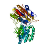
| ||||||||
|---|---|---|---|---|---|---|---|---|---|
| 1 |
| ||||||||
| Unit cell |
| ||||||||
| Components on special symmetry positions |
| ||||||||
| Details | The biological assembly is a monomer in one asymmetric unit which contains chain A and two Mn ions. |
-Components
| #1: Protein | Mass: 57023.562 Da / Num. of mol.: 1 Source method: isolated from a genetically manipulated source Source: (gene. exp.) Geobacillus stearothermophilus (bacteria)Plasmid: PGS3 / Production host: Bacillus subtilis (bacteria) / References: UniProt: Q9X519, EC: 5.4.2.1 | ||||
|---|---|---|---|---|---|
| #2: Chemical |   Mass: 54.938 Da / Num. of mol.: 2 / Source method: obtained synthetically / Formula: Mn Mass: 54.938 Da / Num. of mol.: 2 / Source method: obtained synthetically / Formula: Mn#3: Chemical | ChemComp-3PG / | 3-Phosphoglyceric acid  Mass: 186.057 Da / Num. of mol.: 1 / Source method: obtained synthetically / Formula: C3H7O7P Mass: 186.057 Da / Num. of mol.: 1 / Source method: obtained synthetically / Formula: C3H7O7P#4: Water | ChemComp-HOH / | Water Mass: 18.015 Da / Num. of mol.: 180 / Source method: isolated from a natural source / Formula: H2O Mass: 18.015 Da / Num. of mol.: 180 / Source method: isolated from a natural source / Formula: H2O |
-Experimental details
-Experiment
| Experiment | Method: X-RAY DIFFRACTION / Number of used crystals: 3 |
|---|
- Sample preparation
| Crystal | Density Matthews: 3.31 Å3/Da / Density % sol: 62.82 % | ||||||||||||||||||||||||||||||||||||||||||||||||||||||
|---|---|---|---|---|---|---|---|---|---|---|---|---|---|---|---|---|---|---|---|---|---|---|---|---|---|---|---|---|---|---|---|---|---|---|---|---|---|---|---|---|---|---|---|---|---|---|---|---|---|---|---|---|---|---|---|
| Crystal grow | Temperature: 295 K / Method: vapor diffusion, hanging drop / pH: 7.4 Details: 2.0 M ammonium sulfate, 25 mM zinc acetate, 20 mM cesium chloride, 15 mM 2-mercaptoethanol, 3% polyethylene glycol 200, 50 mM Tris-HCl (pH7.4) , VAPOR DIFFUSION, HANGING DROP, temperature 295K | ||||||||||||||||||||||||||||||||||||||||||||||||||||||
| Crystal grow | *PLUS Temperature: 22 ℃ / Details: Chander, M., (1999) J.Struct.Biol., 126, 156. | ||||||||||||||||||||||||||||||||||||||||||||||||||||||
| Components of the solutions | *PLUS
|
-Data collection
| Diffraction | Mean temperature: 100 K |
|---|---|
| Diffraction source | Source: SYNCHROTRON / Site: NSLS  / Beamline: X25 / Wavelength: 1.1 / Beamline: X25 / Wavelength: 1.1 |
| Detector | Type: BRANDEIS - B4 / Detector: CCD / Date: Feb 11, 1999 |
| Radiation | Protocol: SINGLE WAVELENGTH / Monochromatic (M) / Laue (L): M / Scattering type: x-ray |
| Radiation wavelength | Wavelength: 1.1 Å / Relative weight: 1 |
| Reflection | Resolution: 1.9→50 Å / Num. all: 57256 / Num. obs: 57256 / % possible obs: 96.2 % / Observed criterion σ(F): 2 / Observed criterion σ(I): 2 / Redundancy: 3.8 % / Rmerge(I) obs: 0.099 / Net I/σ(I): 6.6 |
| Reflection shell | Resolution: 1.9→1.97 Å / Redundancy: 2.1 % / Rmerge(I) obs: 0.233 / Num. unique all: 4983 / % possible all: 85.1 |
| Reflection | *PLUS |
| Reflection shell | *PLUS % possible obs: 85.1 % |
- Processing
| Software |
| ||||||||||||||||||||||||||||||
|---|---|---|---|---|---|---|---|---|---|---|---|---|---|---|---|---|---|---|---|---|---|---|---|---|---|---|---|---|---|---|---|
| Refinement | Resolution: 1.9→20 Å / σ(F): 2 / σ(I): 0.001 / Stereochemistry target values: Engh & Huber / Details: X-plor simulated annealing
| ||||||||||||||||||||||||||||||
| Refinement step | Cycle: LAST / Resolution: 1.9→20 Å
| ||||||||||||||||||||||||||||||
| Software | *PLUS Name: X-PLOR / Version: 3.851 / Classification: refinement | ||||||||||||||||||||||||||||||
| Refinement | *PLUS Lowest resolution: 30 Å / σ(F): 2 / Rfactor obs: 0.2071 / Rfactor Rfree: 0.247 / % reflection Rfree: 10 % | ||||||||||||||||||||||||||||||
| Solvent computation | *PLUS | ||||||||||||||||||||||||||||||
| Displacement parameters | *PLUS | ||||||||||||||||||||||||||||||
| Refine LS restraints | *PLUS
| ||||||||||||||||||||||||||||||
| LS refinement shell | *PLUS Rfactor Rfree: 0.3476 / Rfactor obs: 0.365 |
