 Movie
Movie Controller

[English] 日本語
 Yorodumi
- PDB-1eix: STRUCTURE OF OROTIDINE 5'-MONOPHOSPHATE DECARBOXYLASE FROM E. COL... -
+ Open data

- Basic information
| Entry | Database: PDB / ID: 1eix | ||||||
|---|---|---|---|---|---|---|---|
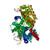
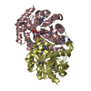
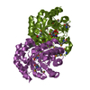
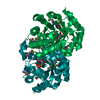
| Title | STRUCTURE OF OROTIDINE 5'-MONOPHOSPHATE DECARBOXYLASE FROM E. COLI, CO-CRYSTALLISED WITH THE INHIBITOR BMP | ||||||
 Components Components | OROTIDINE 5'-MONOPHOSPHATE DECARBOXYLASE | ||||||
 Keywords Keywords |  LYASE / alpha-beta-barrel / protein-inhibitor complex / homodimer LYASE / alpha-beta-barrel / protein-inhibitor complex / homodimer | ||||||
| Function / homology |  Function and homology information Function and homology informationnucleobase-containing small molecule interconversion / orotidine-5'-phosphate decarboxylase / orotidine-5'-phosphate decarboxylase activity / carboxy-lyase activity / 'de novo' UMP biosynthetic process / 'de novo' pyrimidine nucleobase biosynthetic process / cytosol / cytoplasmSimilarity search - Function | ||||||
| Biological species |  Escherichia coli (E. coli) Escherichia coli (E. coli) | ||||||
| Method | X-RAY DIFFRACTION / SYNCHROTRON / Resolution: 2.5 Å | ||||||
 Authors Authors | Harris, P. / Poulsen, J.C.N. / Jensen, K.F. / Larsen, S. | ||||||
 Citation Citation | Journal: Biochemistry / Year: 2000 Title: Structural basis for the catalytic mechanism of a proficient enzyme: orotidine 5'-monophosphate decarboxylase. Authors: Harris, P. / Poulsen, J.C.N. / Jensen, K.F. / Larsen, S. | ||||||
| History |
|
- Structure visualization
| Structure viewer | Molecule: MolmilJmol/JSmol |
|---|
- Downloads & links
-Download
| PDBx/mmCIF format | 1eix.cif.gz | 189.5 KB | Display | PDBx/mmCIF format |
|---|---|---|---|---|
| PDB format | pdb1eix.ent.gz | 152.3 KB | Display | PDB format |
| PDBx/mmJSON format | 1eix.json.gz | Tree view | PDBx/mmJSON format | |
| Others |  Other downloads Other downloads |
-Validation report
| Arichive directory | https://data.pdbj.org/pub/pdb/validation_reports/ei/1eixftp://data.pdbj.org/pub/pdb/validation_reports/ei/1eix | HTTPS FTP |
|---|
-Related structure data
| Similar structure data |
|---|


-Links
 PDBj
PDBj
- Assembly
| Deposited unit | 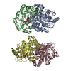
| ||||||||
|---|---|---|---|---|---|---|---|---|---|
| 1 | 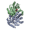
| ||||||||
| 2 | 
| ||||||||
| Unit cell |
| ||||||||
| Details | The biological assembly is a dimer constructed from chain A and B (or C and D) which are connected by a twofold. |
-Components
| #1: Protein | Mass: 26378.225 Da / Num. of mol.: 4 Source method: isolated from a genetically manipulated source Source: (gene. exp.) Escherichia coli (E. coli) / Plasmid: PLFF8 / Production host: Escherichia coli (E. coli)References: UniProt: P08244, orotidine-5'-phosphate decarboxylase#2: Chemical | ChemComp-BMQ /   Type: RNA linking / Mass: 340.181 Da / Num. of mol.: 4 / Source method: obtained synthetically / Formula: C9H13N2O10P / Details: SIGMA Type: RNA linking / Mass: 340.181 Da / Num. of mol.: 4 / Source method: obtained synthetically / Formula: C9H13N2O10P / Details: SIGMA#3: Water | ChemComp-HOH / | Water Mass: 18.015 Da / Num. of mol.: 379 / Source method: isolated from a natural source / Formula: H2O Mass: 18.015 Da / Num. of mol.: 379 / Source method: isolated from a natural source / Formula: H2O |
|---|
-Experimental details
-Experiment
| Experiment | Method: X-RAY DIFFRACTION / Number of used crystals: 1 |
|---|
- Sample preparation
| Crystal | Density Matthews: 2.02 Å3/Da / Density % sol: 39.07 % | ||||||||||||||||||||||||
|---|---|---|---|---|---|---|---|---|---|---|---|---|---|---|---|---|---|---|---|---|---|---|---|---|---|
| Crystal grow | Temperature: 290 K / Method: vapor diffusion, hanging drop / pH: 7 Details: PEG 8000, magnesium chloride, pH 7.0, VAPOR DIFFUSION, HANGING DROP, temperature 290.0K | ||||||||||||||||||||||||
| Crystal grow | *PLUS Temperature: 17 ℃ | ||||||||||||||||||||||||
| Components of the solutions | *PLUS
|
-Data collection
| Diffraction | Mean temperature: 100 K |
|---|---|
| Diffraction source | Source: SYNCHROTRON / Site: MAX II  / Beamline: I711 / Wavelength: 0.9902 / Beamline: I711 / Wavelength: 0.9902 |
| Detector | Type: MARRESEARCH / Detector: IMAGE PLATE / Date: Apr 26, 1999 |
| Radiation | Protocol: SINGLE WAVELENGTH / Monochromatic (M) / Laue (L): M / Scattering type: x-ray |
| Radiation wavelength | Wavelength: 0.9902 Å / Relative weight: 1 |
| Reflection | Resolution: 2.5→30 Å / Num. all: 102335 / Num. obs: 29678 / % possible obs: 97.7 % / Observed criterion σ(F): 0 / Observed criterion σ(I): 0 / Redundancy: 3.4 % / Biso Wilson estimate: 18.2 Å2 / Rmerge(I) obs: 0.088 / Net I/σ(I): 10.6 |
| Reflection shell | Resolution: 2.5→2.64 Å / Redundancy: 3.3 % / Rmerge(I) obs: 0.243 / Num. unique all: 4946 / % possible all: 98 |
| Reflection | *PLUS Num. measured all: 102335 |
| Reflection shell | *PLUS Highest resolution: 2.5 Å / % possible obs: 92.4 % / Mean I/σ(I) obs: 5.2 |
- Processing
| Software |
| |||||||||||||||||||||||||
|---|---|---|---|---|---|---|---|---|---|---|---|---|---|---|---|---|---|---|---|---|---|---|---|---|---|---|
| Refinement | Resolution: 2.5→30 Å / σ(F): 0 / σ(I): 0 / Stereochemistry target values: Engh & Huber
| |||||||||||||||||||||||||
| Refinement step | Cycle: LAST / Resolution: 2.5→30 Å
| |||||||||||||||||||||||||
| Refine LS restraints |
| |||||||||||||||||||||||||
| Software | *PLUS Name: CNS / Classification: refinement | |||||||||||||||||||||||||
| Refinement | *PLUS Highest resolution: 2.5 Å / Lowest resolution: 30 Å / σ(F): 0 / Rfactor obs: 0.217 | |||||||||||||||||||||||||
| Solvent computation | *PLUS | |||||||||||||||||||||||||
| Displacement parameters | *PLUS | |||||||||||||||||||||||||
| Refine LS restraints | *PLUS Type: c_angle_deg / Dev ideal: 1.3 |
