 Movie
Movie Controller

[English] 日本語
 Yorodumi
- PDB-5eb8: Crystal structure of aromatic mutant (F4W) of an alkali thermosta... -
+ Open data

- Basic information
| Entry | Database: PDB / ID: 5eb8 | ||||||
|---|---|---|---|---|---|---|---|
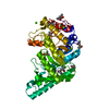
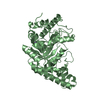
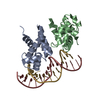
| Title | Crystal structure of aromatic mutant (F4W) of an alkali thermostable GH10 xylanase from Bacillus sp. NG-27 | ||||||
 Components Components | Beta-xylanase Xylanase Xylanase | ||||||
 Keywords Keywords | HYDROLASE / glycosyl hydrolase famility 10 / xylanase / (beta/alpha)8-barrel | ||||||
| Function / homology |  Function and homology informationendo-1,4-beta-xylanase activity / endo-1,4-beta-xylanase / xylan catabolic process / metal ion binding Function and homology informationendo-1,4-beta-xylanase activity / endo-1,4-beta-xylanase / xylan catabolic process / metal ion bindingSimilarity search - Function | ||||||
| Biological species |  | ||||||
| Method | X-RAY DIFFRACTION / MOLECULAR REPLACEMENT / Resolution: 2.22 Å | ||||||
 Authors Authors | Mahanta, P. / Bhardwaj, A. / Reddy, V.S. / Ramakumar, S. | ||||||
 Citation Citation | Journal: To Be Published Title: Crystal structure of aromatic mutant (F4W) of an alkali thermostable GH10 xylanase from Bacillus sp. NG-27 Authors: Mahanta, P. / Bhardwaj, A. / Reddy, V.S. / Ramakumar, S. | ||||||
| History |
|
- Structure visualization
| Structure viewer | Molecule: MolmilJmol/JSmol |
|---|
- Downloads & links
-Download
| PDBx/mmCIF format | 5eb8.cif.gz | 302.7 KB | Display | PDBx/mmCIF format |
|---|---|---|---|---|
| PDB format | pdb5eb8.ent.gz | 244.9 KB | Display | PDB format |
| PDBx/mmJSON format | 5eb8.json.gz | Tree view | PDBx/mmJSON format | |
| Others |  Other downloads Other downloads |
-Validation report
| Arichive directory | https://data.pdbj.org/pub/pdb/validation_reports/eb/5eb8ftp://data.pdbj.org/pub/pdb/validation_reports/eb/5eb8 | HTTPS FTP |
|---|
-Related structure data
| Related structure data |  2f8qS S: Starting model for refinement |
|---|---|
| Similar structure data |


-Links
 PDBj
PDBj
- Assembly
| Deposited unit | 
| ||||||||
|---|---|---|---|---|---|---|---|---|---|
| 1 | 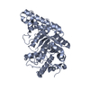
| ||||||||
| 2 | 
| ||||||||
| Unit cell |
|
-Components
| #1: Protein | Xylanase / alkali thermostable GH10 endoxylanase Mass: 41152.477 Da / Num. of mol.: 2 / Fragment: UNP residues 52-405 / Mutation: F4W Source method: isolated from a genetically manipulated source Source: (gene. exp.) Production host: Escherichia coli 'BL21-Gold(DE3)pLysS AG' (bacteria)Strain (production host): BL21-Gold(DE3)pLysS AG / References: UniProt: O30700, endo-1,4-beta-xylanase#2: Chemical |   Mass: 24.305 Da / Num. of mol.: 2 / Source method: obtained synthetically / Formula: Mg Mass: 24.305 Da / Num. of mol.: 2 / Source method: obtained synthetically / Formula: Mg#3: Chemical |   Mass: 22.990 Da / Num. of mol.: 2 / Source method: obtained synthetically / Formula: Na Mass: 22.990 Da / Num. of mol.: 2 / Source method: obtained synthetically / Formula: Na#4: Water | ChemComp-HOH / | Water Mass: 18.015 Da / Num. of mol.: 417 / Source method: isolated from a natural source / Formula: H2O Mass: 18.015 Da / Num. of mol.: 417 / Source method: isolated from a natural source / Formula: H2O |
|---|
-Experimental details
-Experiment
| Experiment | Method: X-RAY DIFFRACTION / Number of used crystals: 1 |
|---|
- Sample preparation
| Crystal | Density Matthews: 2.3 Å3/Da / Density % sol: 46.56 % / Description: Plate shaped |
|---|---|
| Crystal grow | Temperature: 293 K / Method: microbatch / pH: 8.5 Details: 0.1M NaCl, 120mM MgCl2, 0.1M Tris-HCl pH 8.5, 18% PEG 8000 |
-Data collection
| Diffraction | Mean temperature: 100 K | ||||||||||||||||||||||||||||||||||||||||||||||||||||||||||||||||||||||||||||||||||||||||||||||||||||||||||||||
|---|---|---|---|---|---|---|---|---|---|---|---|---|---|---|---|---|---|---|---|---|---|---|---|---|---|---|---|---|---|---|---|---|---|---|---|---|---|---|---|---|---|---|---|---|---|---|---|---|---|---|---|---|---|---|---|---|---|---|---|---|---|---|---|---|---|---|---|---|---|---|---|---|---|---|---|---|---|---|---|---|---|---|---|---|---|---|---|---|---|---|---|---|---|---|---|---|---|---|---|---|---|---|---|---|---|---|---|---|---|---|---|
| Diffraction source | Source: ROTATING ANODE / Type: RIGAKU ULTRAX 18 / Wavelength: 1.54179 Å | ||||||||||||||||||||||||||||||||||||||||||||||||||||||||||||||||||||||||||||||||||||||||||||||||||||||||||||||
| Detector | Type: RIGAKU / Detector: IMAGE PLATE / Date: Jun 6, 2012 | ||||||||||||||||||||||||||||||||||||||||||||||||||||||||||||||||||||||||||||||||||||||||||||||||||||||||||||||
| Radiation | Protocol: SINGLE WAVELENGTH / Monochromatic (M) / Laue (L): M / Scattering type: x-ray | ||||||||||||||||||||||||||||||||||||||||||||||||||||||||||||||||||||||||||||||||||||||||||||||||||||||||||||||
| Radiation wavelength | Wavelength: 1.54179 Å / Relative weight: 1 | ||||||||||||||||||||||||||||||||||||||||||||||||||||||||||||||||||||||||||||||||||||||||||||||||||||||||||||||
| Reflection | Resolution: 2.219→88.375 Å / Num. all: 38071 / Num. obs: 38071 / % possible obs: 98.4 % / Redundancy: 7.1 % / Rpim(I) all: 0.033 / Rrim(I) all: 0.089 / Rsym value: 0.083 / Net I/av σ(I): 6.565 / Net I/σ(I): 15.1 / Num. measured all: 270609 | ||||||||||||||||||||||||||||||||||||||||||||||||||||||||||||||||||||||||||||||||||||||||||||||||||||||||||||||
| Reflection shell | Diffraction-ID: 1 / Rejects: 0
|
- Processing
| Software |
| |||||||||||||||||||||||||||||||||||||||||||||||||||||||||||||||||||||||||||
|---|---|---|---|---|---|---|---|---|---|---|---|---|---|---|---|---|---|---|---|---|---|---|---|---|---|---|---|---|---|---|---|---|---|---|---|---|---|---|---|---|---|---|---|---|---|---|---|---|---|---|---|---|---|---|---|---|---|---|---|---|---|---|---|---|---|---|---|---|---|---|---|---|---|---|---|---|
| Refinement | Method to determine structure: MOLECULAR REPLACEMENT Starting model: 2F8Q Resolution: 2.22→88.375 Å / Cor.coef. Fo:Fc: 0.955 / Cor.coef. Fo:Fc free: 0.931 / WRfactor Rfree: 0.2111 / WRfactor Rwork: 0.166 / FOM work R set: 0.8672 / SU B: 9.65 / SU ML: 0.131 / SU R Cruickshank DPI: 0.2827 / SU Rfree: 0.2007 / Cross valid method: THROUGHOUT / σ(F): 0 / ESU R: 0.283 / ESU R Free: 0.201 / Stereochemistry target values: MAXIMUM LIKELIHOOD Details: HYDROGENS HAVE BEEN ADDED IN THE RIDING POSITIONS U VALUES : WITH TLS ADDED
| |||||||||||||||||||||||||||||||||||||||||||||||||||||||||||||||||||||||||||
| Solvent computation | Ion probe radii: 0.8 Å / Shrinkage radii: 0.8 Å / VDW probe radii: 1.2 Å / Solvent model: MASK | |||||||||||||||||||||||||||||||||||||||||||||||||||||||||||||||||||||||||||
| Displacement parameters | Biso max: 72.4 Å2 / Biso mean: 27.172 Å2 / Biso min: 10.01 Å2
| |||||||||||||||||||||||||||||||||||||||||||||||||||||||||||||||||||||||||||
| Refinement step | Cycle: final / Resolution: 2.22→88.375 Å
| |||||||||||||||||||||||||||||||||||||||||||||||||||||||||||||||||||||||||||
| Refine LS restraints |
| |||||||||||||||||||||||||||||||||||||||||||||||||||||||||||||||||||||||||||
| LS refinement shell | Resolution: 2.219→2.277 Å / Total num. of bins used: 20
| |||||||||||||||||||||||||||||||||||||||||||||||||||||||||||||||||||||||||||
| Refinement TLS params. | Method: refined / Refine-ID: X-RAY DIFFRACTION
| |||||||||||||||||||||||||||||||||||||||||||||||||||||||||||||||||||||||||||
| Refinement TLS group |
|
