 Movie
Movie Controller

[English] 日本語
 Yorodumi
- PDB-1e8u: Structure of the multifunctional paramyxovirus hemagglutinin-neur... -
+ Open data

- Basic information
| Entry | Database: PDB / ID: 1e8u | ||||||
|---|---|---|---|---|---|---|---|
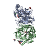
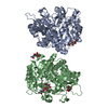
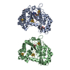
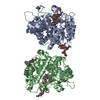
| Title | Structure of the multifunctional paramyxovirus hemagglutinin-neuraminidase | ||||||
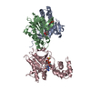
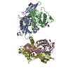
 Components Components | HEMAGGLUTININ-NEURAMINIDASE | ||||||
 Keywords Keywords | HYDROLASE / SIALIDASE / NEURAMINIDASE / HEMAGGLUTININ | ||||||
| Function / homology |  Function and homology information Function and homology informationexo-alpha-(2->3)-sialidase activity / exo-alpha-(2->6)-sialidase activity / exo-alpha-(2->8)-sialidase activity / exo-alpha-sialidase / host cell surface receptor binding / symbiont entry into host cell / viral envelope / virion attachment to host cell / host cell plasma membrane / virion membrane / membraneSimilarity search - Function | ||||||
| Biological species |  NEWCASTLE DISEASE VIRUS NEWCASTLE DISEASE VIRUS | ||||||
| Method | X-RAY DIFFRACTION / SYNCHROTRON / MOLECULAR REPLACEMENT / Resolution: 2 Å | ||||||
 Authors Authors | Crennell, S. / Takimoto, T. / Portner, A. / Taylor, G. | ||||||
 Citation Citation | Journal: Nat.Struct.Biol. / Year: 2000 Title: Crystal Structure of the Multifunctional Paramyxovirus Hemagglutinin-Neuraminidase Authors: Crennell, S. / Takimoto, T. / Portner, A. / Taylor, G. #1: Journal: Virology / Year: 2000 Title: Crystallization of Newcastle Disease Virus Hemagglutinin-Neuraminidase Glycoprotein Authors: Takimoto, T. / Taylor, G.L. / Crennell, S.J. / Scroggs, R.A. / Portner, A. | ||||||
| History |
|
- Structure visualization
| Structure viewer | Molecule: MolmilJmol/JSmol |
|---|
- Downloads & links
-Download
| PDBx/mmCIF format | 1e8u.cif.gz | 187.8 KB | Display | PDBx/mmCIF format |
|---|---|---|---|---|
| PDB format | pdb1e8u.ent.gz | 152.7 KB | Display | PDB format |
| PDBx/mmJSON format | 1e8u.json.gz | Tree view | PDBx/mmJSON format | |
| Others |  Other downloads Other downloads |
-Validation report
| Arichive directory | https://data.pdbj.org/pub/pdb/validation_reports/e8/1e8uftp://data.pdbj.org/pub/pdb/validation_reports/e8/1e8u | HTTPS FTP |
|---|
-Related structure data


-Links
 PDBj
PDBj


- Assembly
| Deposited unit | 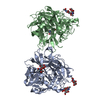
| ||||||||
|---|---|---|---|---|---|---|---|---|---|
| 1 |
| ||||||||
| Unit cell |
| ||||||||
| Noncrystallographic symmetry (NCS) | NCS oper: (Code: given Matrix: (-0.9883, 0.1436, -0.052), Vector : |
-Components
| #1: Protein | / HN Mass: 49856.910 Da / Num. of mol.: 2 / Fragment: HEAD DOMAIN, RESIDUES 124-577 / Source method: isolated from a natural source / Source: (natural) NEWCASTLE DISEASE VIRUS / Strain: KANSAS / References: UniProt: Q9Q2W5, exo-alpha-sialidase#2: Chemical |   Mass: 40.078 Da / Num. of mol.: 2 / Source method: obtained synthetically / Formula: Ca Mass: 40.078 Da / Num. of mol.: 2 / Source method: obtained synthetically / Formula: Ca#3: Sugar | N-Acetylneuraminic acid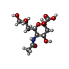  Type: D-saccharide, beta linking / Mass: 309.270 Da / Num. of mol.: 2 Type: D-saccharide, beta linking / Mass: 309.270 Da / Num. of mol.: 2Source method: isolated from a genetically manipulated source Formula: C11H19NO9 #4: Sugar | ChemComp-NAG / N-Acetylglucosamine  Type: D-saccharide, beta linking / Mass: 221.208 Da / Num. of mol.: 5 Type: D-saccharide, beta linking / Mass: 221.208 Da / Num. of mol.: 5Source method: isolated from a genetically manipulated source Formula: C8H15NO6 #5: Water | ChemComp-HOH / | Water Mass: 18.015 Da / Num. of mol.: 370 / Source method: isolated from a natural source / Formula: H2O Mass: 18.015 Da / Num. of mol.: 370 / Source method: isolated from a natural source / Formula: H2O |
|---|
-Experimental details
-Experiment
| Experiment | Method: X-RAY DIFFRACTION |
|---|
- Sample preparation
| Crystal | Density Matthews: 2.85 Å3/Da / Density % sol: 56.91 % |
|---|---|
| Crystal grow | pH: 4.6 / Details: pH 4.60 |
-Data collection
| Diffraction | Mean temperature: 100 K |
|---|---|
| Diffraction source | Source: SYNCHROTRON / Site: EMBL/DESY, HAMBURG  / Beamline: X11 / Wavelength: 0.934 / Beamline: X11 / Wavelength: 0.934 |
| Detector | Detector: IMAGE PLATE |
| Radiation | Protocol: SINGLE WAVELENGTH / Monochromatic (M) / Laue (L): M / Scattering type: x-ray |
| Radiation wavelength | Wavelength: 0.934 Å / Relative weight: 1 |
| Reflection | Resolution: 2→30 Å / Num. obs: 75784 / % possible obs: 97 % / Redundancy: 3.7 % / Rmerge(I) obs: 0.071 / Rsym value: 0.071 |
| Reflection | *PLUS Num. measured all: 282836 |
| Reflection shell | *PLUS % possible obs: 98 % / Rmerge(I) obs: 0.258 |
- Processing
| Software | Name: CNS / Classification: refinement | ||||||||||||||||||||||||||||||||||||||||||||||||||||||||||||
|---|---|---|---|---|---|---|---|---|---|---|---|---|---|---|---|---|---|---|---|---|---|---|---|---|---|---|---|---|---|---|---|---|---|---|---|---|---|---|---|---|---|---|---|---|---|---|---|---|---|---|---|---|---|---|---|---|---|---|---|---|---|
| Refinement | Method to determine structure: MOLECULAR REPLACEMENT / Resolution: 2→6 Å / Cross valid method: THROUGHOUT / σ(F): 0
| ||||||||||||||||||||||||||||||||||||||||||||||||||||||||||||
| Refinement step | Cycle: LAST / Resolution: 2→6 Å
| ||||||||||||||||||||||||||||||||||||||||||||||||||||||||||||
| Refine LS restraints |
|
