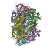
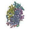
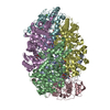
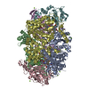
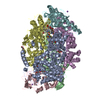
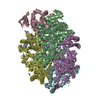
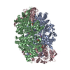
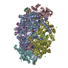
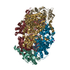
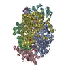
A: METHYL-COENZYME M REDUCTASE I ALPHA SUBUNIT B: METHYL-COENZYME M REDUCTASE I BETA SUBUNIT C: METHYL-COENZYME M REDUCTASE I GAMMA SUBUNIT D: METHYL-COENZYME M REDUCTASE I ALPHA SUBUNIT E: METHYL-COENZYME M REDUCTASE I BETA SUBUNIT F: METHYL-COENZYME M REDUCTASE I GAMMA SUBUNIT hetero molecules
Mass: 142.197 Da / Num. of mol.: 2 / Source method: obtained synthetically / Formula: C2H6O3S2
-
Details
Compound details
THE HEXAMER OF TWO ALPHA, TWO BETA, AND TWO GAMMA CHAINS CATALYZES THE FINAL STEP IN ...THE HEXAMER OF TWO ALPHA, TWO BETA, AND TWO GAMMA CHAINS CATALYZES THE FINAL STEP IN METHANOGENESIS, WHICH IS THE TERMINAL STEP OF ANAEROBIC DEGRADATION OF BIOMASS
-
Experimental details
-
Experiment
Experiment
Method: X-RAY DIFFRACTION / Number of used crystals: 1
-
Sample preparation
Crystal
Density Matthews: 2.24 Å3/Da / Density % sol: 39 %
Crystal grow
pH: 7 / Details: pH 7.00
Crystal grow
*PLUS
Temperature: 14 ℃ / Method: vapor diffusion, hanging drop
Resolution: 2.7→30 Å / Rfactor Rfree error: 0.006 / Data cutoff high absF: 274750.55 / Isotropic thermal model: GROUP / Cross valid method: THROUGHOUT / σ(F): 0 Details: BULK SOLVENT MODEL USED, REFINEMENT WAS CARRIED WITH NCS RESTRAINTS AND THE PDB GENERATED THE ATOMS FOR CHAINS D, E, F. MCR FROM M. KANDLERI MIGHT CONTAIN MODIFIED AMINO ACIDS ANALOGOUS TO ...Details: BULK SOLVENT MODEL USED, REFINEMENT WAS CARRIED WITH NCS RESTRAINTS AND THE PDB GENERATED THE ATOMS FOR CHAINS D, E, F. MCR FROM M. KANDLERI MIGHT CONTAIN MODIFIED AMINO ACIDS ANALOGOUS TO MCR FROM M. THERMOAUTOTROPHICUM AND M. BARKERI. THE DATA SET IS OF UNUSUALLY LOW COMPLETENESS CORRESPONDING TO ABOUT 3.2 A EFFECTIVE RESOLUTION.
Rfactor
Num. reflection
% reflection
Selection details
Rfree
0.278
2196
5 %
RANDOM
Rwork
0.239
-
-
-
obs
0.239
43932
62.9 %
-
Displacement parameters
Biso mean: 25.5 Å2
Baniso -1
Baniso -2
Baniso -3
1-
-9.3 Å2
0 Å2
0 Å2
2-
-
-2.84 Å2
0 Å2
3-
-
-
12.14 Å2
Refine analyze
Free
Obs
Luzzati coordinate error
0.42 Å
0.36 Å
Luzzati d res low
-
5 Å
Luzzati sigma a
0.46 Å
0.36 Å
Refinement step
Cycle: LAST / Resolution: 2.7→30 Å
Protein
Nucleic acid
Ligand
Solvent
Total
Num. atoms
19222
0
180
0
19402
Refine LS restraints
Refine-ID
Type
Dev ideal
X-RAY DIFFRACTION
c_bond_d
0.005
X-RAY DIFFRACTION
c_bond_d_na
X-RAY DIFFRACTION
c_bond_d_prot
X-RAY DIFFRACTION
c_angle_d
X-RAY DIFFRACTION
c_angle_d_na
X-RAY DIFFRACTION
c_angle_d_prot
X-RAY DIFFRACTION
c_angle_deg
0.8
X-RAY DIFFRACTION
c_angle_deg_na
X-RAY DIFFRACTION
c_angle_deg_prot
X-RAY DIFFRACTION
c_dihedral_angle_d
24.8
X-RAY DIFFRACTION
c_dihedral_angle_d_na
X-RAY DIFFRACTION
c_dihedral_angle_d_prot
X-RAY DIFFRACTION
c_improper_angle_d
1.55
X-RAY DIFFRACTION
c_improper_angle_d_na
X-RAY DIFFRACTION
c_improper_angle_d_prot
X-RAY DIFFRACTION
c_mcbond_it
X-RAY DIFFRACTION
c_mcangle_it
X-RAY DIFFRACTION
c_scbond_it
X-RAY DIFFRACTION
c_scangle_it
Refine LS restraints NCS
NCS model details: CONSTR
LS refinement shell
Resolution: 2.7→2.87 Å / Rfactor Rfree error: 0.019 / Total num. of bins used: 6
In the structure databanks used in Yorodumi, some data are registered as the other names, "COVID-19 virus" and "2019-nCoV". Here are the details of the virus and the list of structure data.
Jan 31, 2019. EMDB accession codes are about to change! (news from PDBe EMDB page)
EMDB accession codes are about to change! (news from PDBe EMDB page)
The allocation of 4 digits for EMDB accession codes will soon come to an end. Whilst these codes will remain in use, new EMDB accession codes will include an additional digit and will expand incrementally as the available range of codes is exhausted. The current 4-digit format prefixed with “EMD-” (i.e. EMD-XXXX) will advance to a 5-digit format (i.e. EMD-XXXXX), and so on. It is currently estimated that the 4-digit codes will be depleted around Spring 2019, at which point the 5-digit format will come into force.
The EM Navigator/Yorodumi systems omit the EMD- prefix.
Related info.:Q: What is EMD? / ID/Accession-code notation in Yorodumi/EM Navigator
Yorodumi is a browser for structure data from EMDB, PDB, SASBDB, etc.
This page is also the successor to EM Navigator detail page, and also detail information page/front-end page for Omokage search.
The word "yorodu" (or yorozu) is an old Japanese word meaning "ten thousand". "mi" (miru) is to see.
Related info.:EMDB / PDB / SASBDB / Comparison of 3 databanks / Yorodumi Search / Aug 31, 2016. New EM Navigator & Yorodumi / Yorodumi Papers / Jmol/JSmol / Function and homology information / Changes in new EM Navigator and Yorodumi
 Movie
Movie Controller


 Open data
 Basic information
 Components
 Coenzyme-B sulfoethylthiotransferase) x 3
  Keywords
 Function and homology information

 Authors
 Citation
 Structure visualization
 Downloads & links
 Other downloads

 PDBj
PDBj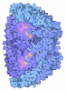
 Assembly
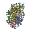
 Mass: 906.580 Da / Num. of mol.: 2 / Source method: obtained synthetically / Formula: C42H51N6NiO13
 Mass: 343.334 Da / Num. of mol.: 2 / Source method: obtained synthetically / Formula: C11H22NO7PS
 Mass: 142.197 Da / Num. of mol.: 2 / Source method: obtained synthetically / Formula: C2H6O3S2
 Sample preparation
 / Beamline: BW7B / Wavelength: 0.84
  Processing