 Movie
Movie Controller


+ Open data

- Basic information
| Entry | Database: PDB / ID: 1dsx | ||||||
|---|---|---|---|---|---|---|---|
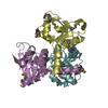
| Title | KV1.2 T1 DOMAIN, RESIDUES 33-119, T46V MUTANT | ||||||
 Components Components | PROTEIN (KV1.2 VOLTAGE-GATED POTASSIUM CHANNEL) | ||||||
 Keywords Keywords |  SIGNALING PROTEIN / VOLTAGE-GATED POTASSIUM CHANNEL / ASSEMBLY DOMAIN / TETRAMER SIGNALING PROTEIN / VOLTAGE-GATED POTASSIUM CHANNEL / ASSEMBLY DOMAIN / TETRAMER | ||||||
| Function / homology |  Function and homology information Function and homology informationoptic nerve structural organization / Voltage gated Potassium channels / voltage-gated monoatomic ion channel activity involved in regulation of postsynaptic membrane potential / potassium channel complex / paranodal junction / potassium ion export across plasma membrane / regulation of circadian sleep/wake cycle, non-REM sleep / axon initial segment / corpus callosum development / voltage-gated monoatomic ion channel activity involved in regulation of presynaptic membrane potential ...optic nerve structural organization / Voltage gated Potassium channels / voltage-gated monoatomic ion channel activity involved in regulation of postsynaptic membrane potential / potassium channel complex / paranodal junction / potassium ion export across plasma membrane / regulation of circadian sleep/wake cycle, non-REM sleep / axon initial segment / corpus callosum development / voltage-gated monoatomic ion channel activity involved in regulation of presynaptic membrane potential / juxtaparanode region of axon / delayed rectifier potassium channel activity / optic nerve development / outward rectifier potassium channel activity / neuronal cell body membrane / regulation of dopamine secretion / voltage-gated potassium channel activity / kinesin binding / lamellipodium membrane / calyx of Held / neuronal action potential / axon terminus / potassium ion transmembrane transport / voltage-gated potassium channel complex / sensory perception of pain / protein homooligomerization / cerebral cortex development / lamellipodium / presynaptic membrane / perikaryon / postsynaptic membrane / endosome / axon / glutamatergic synapse / dendrite / endoplasmic reticulum membrane / plasma membraneSimilarity search - Function | ||||||
| Biological species |  Rattus norvegicus (Norway rat) Rattus norvegicus (Norway rat) | ||||||
| Method | X-RAY DIFFRACTION / SYNCHROTRON / Resolution: 1.6 Å | ||||||
 Authors Authors | Minor Jr., D.L. / Lin, Y.-F. / Mobley, B.C. / Avelar, A. / Jan, Y.N. / Jan, L.Y. / Berger, J.M. | ||||||
 Citation Citation | Journal: Cell(Cambridge,Mass.) / Year: 2000 Title: The polar T1 interface is linked to conformational changes that open the voltage-gated potassium channel. Authors: Minor, D.L. / Lin, Y.F. / Mobley, B.C. / Avelar, A. / Jan, Y.N. / Jan, L.Y. / Berger, J.M. | ||||||
| History |
|
- Structure visualization
| Structure viewer | Molecule: MolmilJmol/JSmol |
|---|
- Downloads & links
-Download
| PDBx/mmCIF format | 1dsx.cif.gz | 161.2 KB | Display | PDBx/mmCIF format |
|---|---|---|---|---|
| PDB format | pdb1dsx.ent.gz | 131.8 KB | Display | PDB format |
| PDBx/mmJSON format | 1dsx.json.gz | Tree view | PDBx/mmJSON format | |
| Others |  Other downloads Other downloads |
-Validation report
| Arichive directory | https://data.pdbj.org/pub/pdb/validation_reports/ds/1dsxftp://data.pdbj.org/pub/pdb/validation_reports/ds/1dsx | HTTPS FTP |
|---|
-Related structure data


-Links
 PDBj
PDBj


- Assembly
| Deposited unit | 
| ||||||||
|---|---|---|---|---|---|---|---|---|---|
| 1 | 
| ||||||||
| 2 | 
| ||||||||
| Unit cell |
|
-Components
| #1: Protein | Mass: 10495.991 Da / Num. of mol.: 8 / Fragment: N-TERMINAL ASSEMBLY DOMAIN, RESIDUES 33-119 / Mutation: YES Source method: isolated from a genetically manipulated source Source: (gene. exp.) Rattus norvegicus (Norway rat) / Organ: BRAIN / Plasmid: PET24 / Production host:  Escherichia coli (E. coli) / References: UniProt: P63142 Escherichia coli (E. coli) / References: UniProt: P63142#2: Water | ChemComp-HOH / | Water Mass: 18.015 Da / Num. of mol.: 551 / Source method: isolated from a natural source / Formula: H2O Mass: 18.015 Da / Num. of mol.: 551 / Source method: isolated from a natural source / Formula: H2O |
|---|
-Experimental details
-Experiment
| Experiment | Method: X-RAY DIFFRACTION / Number of used crystals: 1 |
|---|
- Sample preparation
| Crystal | Density Matthews: 2.26 Å3/Da / Density % sol: 45.62 % | ||||||||||||||||||||||||||||||||||||||||||
|---|---|---|---|---|---|---|---|---|---|---|---|---|---|---|---|---|---|---|---|---|---|---|---|---|---|---|---|---|---|---|---|---|---|---|---|---|---|---|---|---|---|---|---|
| Crystal grow | pH: 8.5 Details: 22% PEG 1500, 5 % ISOPROPANOL, 200 MM NA ACETATE, 12 MM SRCL2, 50 MM TRIS, PH 8.5, pH 8.50 | ||||||||||||||||||||||||||||||||||||||||||
| Crystal grow | *PLUS Temperature: 18 ℃ / pH: 8.5 / Method: vapor diffusion | ||||||||||||||||||||||||||||||||||||||||||
| Components of the solutions | *PLUS
|
-Data collection
| Diffraction | Mean temperature: 77 K |
|---|---|
| Diffraction source | Source: SYNCHROTRON / Site: ALS  / Beamline: 5.0.2 / Wavelength: 1 / Beamline: 5.0.2 / Wavelength: 1 |
| Detector | Type: ADSC / Detector: CCD / Date: Aug 20, 1999 |
| Radiation | Protocol: SINGLE WAVELENGTH / Monochromatic (M) / Laue (L): M / Scattering type: x-ray |
| Radiation wavelength | Wavelength: 1 Å / Relative weight: 1 |
| Reflection | Resolution: 1.6→30 Å / Num. obs: 97159 / % possible obs: 99.8 % / Observed criterion σ(I): 0 / Redundancy: 7.7 % / Biso Wilson estimate: 23.1 Å2 / Rmerge(I) obs: 0.048 / Net I/σ(I): 13.8 |
| Reflection shell | Resolution: 1.6→1.75 Å / Rmerge(I) obs: 0.371 / % possible all: 84.4 |
| Reflection | *PLUS Highest resolution: 1.6 Å / Lowest resolution: 30 Å / % possible obs: 96.1 % / Observed criterion σ(I): 0 / Redundancy: 7.7 % / Biso Wilson estimate: 23.1 Å2 |
- Processing
| Software |
| ||||||||||||||||
|---|---|---|---|---|---|---|---|---|---|---|---|---|---|---|---|---|---|
| Refinement | Resolution: 1.6→30 Å / σ(F): 0
| ||||||||||||||||
| Refinement step | Cycle: LAST / Resolution: 1.6→30 Å
| ||||||||||||||||
| Software | *PLUS Name: REFMAC / Classification: refinement | ||||||||||||||||
| Refinement | *PLUS % reflection Rfree: 10 % / Rfactor obs: 0.234 | ||||||||||||||||
| Solvent computation | *PLUS | ||||||||||||||||
| Displacement parameters | *PLUS | ||||||||||||||||
| Refine LS restraints | *PLUS
|
