 Movie
Movie Controller

[English] 日本語
 Yorodumi
- PDB-3fnj: Crystal structure of the full-length lp_1913 protein from Lactoba... -
+ Open data

- Basic information
| Entry | Database: PDB / ID: 3fnj | ||||||
|---|---|---|---|---|---|---|---|
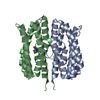
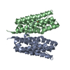
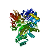
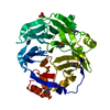
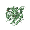
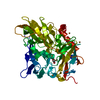
| Title | Crystal structure of the full-length lp_1913 protein from Lactobacillus plantarum, Northeast Structural Genomics Consortium Target LpR140 | ||||||
 Components Components | protein lp_1913 | ||||||
 Keywords Keywords |  structural genomics / unknown function / alpha-beta protein / PSI-2 / Protein Structure Initiative / Northeast Structural Genomics Consortium / NESG structural genomics / unknown function / alpha-beta protein / PSI-2 / Protein Structure Initiative / Northeast Structural Genomics Consortium / NESG | ||||||
| Function / homology |  Function and homology information Function and homology information | ||||||
| Biological species |  Lactobacillus plantarum (bacteria) Lactobacillus plantarum (bacteria) | ||||||
| Method | X-RAY DIFFRACTION / SYNCHROTRON / SAD / Resolution: 2.7 Å | ||||||
 Authors Authors | Seetharaman, J. / Chen, Y. / Forouhar, F. / Sahdev, S. / Janjua, H. / Xiao, R. / Ciccosanti, C. / Foote, E.L. / Acton, T.B. / Rost, B. ...Seetharaman, J. / Chen, Y. / Forouhar, F. / Sahdev, S. / Janjua, H. / Xiao, R. / Ciccosanti, C. / Foote, E.L. / Acton, T.B. / Rost, B. / Montelione, G.T. / Hunt, J.F. / Tong, L. / Northeast Structural Genomics Consortium (NESG) | ||||||
 Citation Citation | Journal: To be Published Title: Crystal structure of the full-length lp_1913 protein from Lactobacillus plantarum, Northeast Structural Genomics Consortium Target LpR140 Authors: Seetharaman, J. / Chen, Y. / Forouhar, F. / Sahdev, S. / Janjua, H. / Xiao, R. / Ciccosanti, C. / Foote, E.L. / Acton, T.B. / Rost, B. / Montelione, G.T. / Hunt, J.F. / Tong, L. | ||||||
| History |
|
- Structure visualization
| Structure viewer | Molecule: MolmilJmol/JSmol |
|---|
- Downloads & links
-Download
| PDBx/mmCIF format | 3fnj.cif.gz | 143 KB | Display | PDBx/mmCIF format |
|---|---|---|---|---|
| PDB format | pdb3fnj.ent.gz | 121.6 KB | Display | PDB format |
| PDBx/mmJSON format | 3fnj.json.gz | Tree view | PDBx/mmJSON format | |
| Others |  Other downloads Other downloads |
-Validation report
| Arichive directory | https://data.pdbj.org/pub/pdb/validation_reports/fn/3fnjftp://data.pdbj.org/pub/pdb/validation_reports/fn/3fnj | HTTPS FTP |
|---|
-Related structure data
| Similar structure data | |
|---|---|
| Other databases |


-Links
 PDBj
PDBj- Assembly
| Deposited unit | 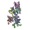
| ||||||||
|---|---|---|---|---|---|---|---|---|---|
| 1 | 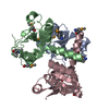
| ||||||||
| 2 | 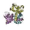
| ||||||||
| Unit cell |
| ||||||||
| Details | trimer |
-Components
| #1: Protein | Mass: 14541.033 Da / Num. of mol.: 6 Source method: isolated from a genetically manipulated source Source: (gene. exp.) Lactobacillus plantarum (bacteria) / Strain: WCFS1 / Gene: lp_1913 / Plasmid: pET21 / Production host: Escherichia coli (E. coli) / Strain (production host): BL21(DE3)+Magic / References: UniProt: Q88VW8, UniProt: F9UPN6*PLUS#2: Water | ChemComp-HOH / | Water Mass: 18.015 Da / Num. of mol.: 82 / Source method: isolated from a natural source / Formula: H2O Mass: 18.015 Da / Num. of mol.: 82 / Source method: isolated from a natural source / Formula: H2O |
|---|
-Experimental details
-Experiment
| Experiment | Method: X-RAY DIFFRACTION / Number of used crystals: 1 |
|---|
- Sample preparation
| Crystal | Density Matthews: 2.77 Å3/Da / Density % sol: 55.56 % |
|---|---|
| Crystal grow | Temperature: 293 K / Method: microbatch under oil / pH: 7 Details: Protein solution: 10 mM Tris (pH 7.5), 100 mM sodium chloride, and 5 mM DTT. Reservoir solution: 100 mM Bis-Tris Propane (pH 7.0) and 1880 mM sodium thiosulfate. , Microbatch under oil, temperature 293K |
-Data collection
| Diffraction | Mean temperature: 100 K |
|---|---|
| Diffraction source | Source: SYNCHROTRON / Site: NSLS  / Beamline: X4A / Wavelength: 0.97905 Å / Beamline: X4A / Wavelength: 0.97905 Å |
| Detector | Type: ADSC QUANTUM 4 / Detector: CCD / Date: Nov 25, 2008 / Details: mirrors |
| Radiation | Monochromator: Si 111 CHANNEL / Protocol: SINGLE WAVELENGTH / Monochromatic (M) / Laue (L): M / Scattering type: x-ray |
| Radiation wavelength | Wavelength: 0.97905 Å / Relative weight: 1 |
| Reflection | Resolution: 2.7→30 Å / Num. all: 51670 / Num. obs: 51464 / % possible obs: 99.6 % / Observed criterion σ(F): 0 / Observed criterion σ(I): 0 / Redundancy: 7.5 % / Biso Wilson estimate: 34.8 Å2 / Rmerge(I) obs: 0.085 / Rsym value: 0.06 / Net I/σ(I): 26.7 |
| Reflection shell | Resolution: 2.7→2.8 Å / Redundancy: 7.3 % / Rmerge(I) obs: 0.421 / Mean I/σ(I) obs: 4.4 / Num. unique all: 5103 / Rsym value: 0.273 / % possible all: 100 |
- Processing
| Software |
| |||||||||||||||||||||||||
|---|---|---|---|---|---|---|---|---|---|---|---|---|---|---|---|---|---|---|---|---|---|---|---|---|---|---|
| Refinement | Method to determine structure: SAD / Resolution: 2.7→19.92 Å / Rfactor Rfree error: 0.007 / Data cutoff high absF: 385035.69 / Data cutoff low absF: 0 / Isotropic thermal model: RESTRAINED / Cross valid method: THROUGHOUT / σ(F): 2 / σ(I): 2 / Stereochemistry target values: Engh & Huber
| |||||||||||||||||||||||||
| Solvent computation | Solvent model: FLAT MODEL / Bsol: 21.0696 Å2 / ksol: 0.3 e/Å3 | |||||||||||||||||||||||||
| Displacement parameters | Biso mean: 55.5 Å2
| |||||||||||||||||||||||||
| Refine analyze |
| |||||||||||||||||||||||||
| Refinement step | Cycle: LAST / Resolution: 2.7→19.92 Å
| |||||||||||||||||||||||||
| Refine LS restraints |
| |||||||||||||||||||||||||
| LS refinement shell | Resolution: 2.7→2.8 Å / Rfactor Rfree error: 0.027 / Total num. of bins used: 10
|
