 Movie
Movie Controller

[English] 日本語
 Yorodumi
- PDB-1dfi: X-RAY STRUCTURE OF ESCHERICHIA COLI ENOYL REDUCTASE WITH BOUND NAD -
+ Open data

- Basic information
| Entry | Database: PDB / ID: 1dfi | ||||||
|---|---|---|---|---|---|---|---|
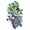
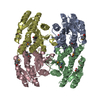
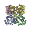
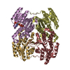
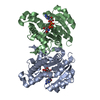
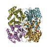
| Title | X-RAY STRUCTURE OF ESCHERICHIA COLI ENOYL REDUCTASE WITH BOUND NAD | ||||||
 Components Components | ENOYL ACYL CARRIER PROTEIN REDUCTASE | ||||||
 Keywords Keywords |  OXIDOREDUCTASE / LIPID BIOSYNTHESIS OXIDOREDUCTASE / LIPID BIOSYNTHESIS | ||||||
| Function / homology |  Function and homology information Function and homology informationenoyl-[acyl-carrier-protein] reductase activity (NAD(P)H) / NADH binding / biotin biosynthetic process / fatty acid elongation / enoyl-[acyl-carrier-protein] reductase (NADH) / enoyl-[acyl-carrier-protein] reductase (NADH) activity / lipid biosynthetic process / catalytic complex / protein homotetramerization / response to antibiotic ...enoyl-[acyl-carrier-protein] reductase activity (NAD(P)H) / NADH binding / biotin biosynthetic process / fatty acid elongation / enoyl-[acyl-carrier-protein] reductase (NADH) / enoyl-[acyl-carrier-protein] reductase (NADH) activity / lipid biosynthetic process / catalytic complex / protein homotetramerization / response to antibiotic / protein-containing complex / membrane / identical protein binding / cytosolSimilarity search - Function | ||||||
| Biological species |  Escherichia coli (E. coli) Escherichia coli (E. coli) | ||||||
| Method | X-RAY DIFFRACTION / SYNCHROTRON / MOLECULAR REPLACEMENT, ISOMORPHOUS REPLACEMENT / Resolution: 2.09 Å | ||||||
 Authors Authors | Baldock, C. / Rafferty, J.B. / Rice, D.W. | ||||||
 Citation Citation | Journal: Science / Year: 1996 Title: A mechanism of drug action revealed by structural studies of enoyl reductase. Authors: Baldock, C. / Rafferty, J.B. / Sedelnikova, S.E. / Baker, P.J. / Stuitje, A.R. / Slabas, A.R. / Hawkes, T.R. / Rice, D.W. #1: Journal: Acta Crystallogr.,Sect.D / Year: 1996Title: Crystallization of Escherichia Coli Enoyl Reductase and its Complex with Diazaborine Authors: Baldock, C. / Rafferty, J.B. / Sedelnikova, S.E. / Bithell, S. / Stuitje, A.R. / Slabas, A.R. / Rice, D.W. | ||||||
| History |
|
- Structure visualization
| Structure viewer | Molecule: MolmilJmol/JSmol |
|---|
- Downloads & links
-Download
| PDBx/mmCIF format | 1dfi.cif.gz | 195.8 KB | Display | PDBx/mmCIF format |
|---|---|---|---|---|
| PDB format | pdb1dfi.ent.gz | 161 KB | Display | PDB format |
| PDBx/mmJSON format | 1dfi.json.gz | Tree view | PDBx/mmJSON format | |
| Others |  Other downloads Other downloads |
-Validation report
| Arichive directory | https://data.pdbj.org/pub/pdb/validation_reports/df/1dfiftp://data.pdbj.org/pub/pdb/validation_reports/df/1dfi | HTTPS FTP |
|---|
-Related structure data
| Related structure data |  1dfgC 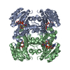 1dfhC 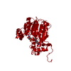 1enoS S: Starting model for refinement C: citing same article ( |
|---|---|
| Similar structure data |


-Links
 PDBj
PDBj

- Assembly
| Deposited unit | 
| ||||||||||||||||
|---|---|---|---|---|---|---|---|---|---|---|---|---|---|---|---|---|---|
| 1 |
| ||||||||||||||||
| Unit cell |
| ||||||||||||||||
| Noncrystallographic symmetry (NCS) | NCS oper:
|
-Components
| #1: Protein | Mass: 27761.730 Da / Num. of mol.: 4 Source method: isolated from a genetically manipulated source Source: (gene. exp.) Escherichia coli (E. coli) / Plasmid: PENVM5 / Species (production host): Escherichia coli / Production host: Escherichia coli BL21 (bacteria) / Strain (production host): BL21References: UniProt: P29132, UniProt: P0AEK4*PLUS, enoyl-[acyl-carrier-protein] reductase (NADH) #2: Chemical | ChemComp-NAD / Nicotinamide adenine dinucleotide  Mass: 663.425 Da / Num. of mol.: 4 / Source method: obtained synthetically / Formula: C21H27N7O14P2 / Comment: NAD*YM Mass: 663.425 Da / Num. of mol.: 4 / Source method: obtained synthetically / Formula: C21H27N7O14P2 / Comment: NAD*YM#3: Water | ChemComp-HOH / | Water Mass: 18.015 Da / Num. of mol.: 327 / Source method: isolated from a natural source / Formula: H2O Mass: 18.015 Da / Num. of mol.: 327 / Source method: isolated from a natural source / Formula: H2O |
|---|
-Experimental details
-Experiment
| Experiment | Method: X-RAY DIFFRACTION / Number of used crystals: 2 |
|---|
- Sample preparation
| Crystal | Density Matthews: 2.11 Å3/Da / Density % sol: 41.7 % | ||||||||||||||||||||||||||||||||||||||||||
|---|---|---|---|---|---|---|---|---|---|---|---|---|---|---|---|---|---|---|---|---|---|---|---|---|---|---|---|---|---|---|---|---|---|---|---|---|---|---|---|---|---|---|---|
| Crystal grow | pH: 5 / Details: 12% PEG 400, PH 5.0 ACETATE, 10MM NAD | ||||||||||||||||||||||||||||||||||||||||||
| Crystal grow | *PLUS pH: 7 / Method: vapor diffusion, hanging dropDetails: Baldock, C., (1996) Acta Crystallogr.,Sect.D, 52, 1181. | ||||||||||||||||||||||||||||||||||||||||||
| Components of the solutions | *PLUS
|
-Data collection
| Diffraction | Mean temperature: 298 K |
|---|---|
| Diffraction source | Source: SYNCHROTRON / Site: SRS  / Beamline: PX7.2 / Wavelength: 1.488 / Beamline: PX7.2 / Wavelength: 1.488 |
| Detector | Type: MARRESEARCH / Detector: IMAGE PLATE / Date: Jul 22, 1995 |
| Radiation | Monochromatic (M) / Laue (L): M / Scattering type: x-ray |
| Radiation wavelength | Wavelength: 1.488 Å / Relative weight: 1 |
| Reflection | Resolution: 2.08→26.35 Å / Num. obs: 51902 / % possible obs: 93.1 % / Redundancy: 2.2 % / Biso Wilson estimate: 33 Å2 / Rmerge(I) obs: 0.057 / Net I/σ(I): 8.2 |
| Reflection shell | Resolution: 2.08→2.14 Å / Redundancy: 1.5 % / Rmerge(I) obs: 0.104 / Mean I/σ(I) obs: 4.6 / % possible all: 71.4 |
| Reflection | *PLUS Num. measured all: 113658 |
- Processing
| Software |
| ||||||||||||||||||||||||||||||||||||||||||||||||||
|---|---|---|---|---|---|---|---|---|---|---|---|---|---|---|---|---|---|---|---|---|---|---|---|---|---|---|---|---|---|---|---|---|---|---|---|---|---|---|---|---|---|---|---|---|---|---|---|---|---|---|---|
| Refinement | Method to determine structure: MOLECULAR REPLACEMENT, ISOMORPHOUS REPLACEMENT Starting model: BRASSICA NAPUS ENR (PDB ENTRY 1ENO) Resolution: 2.09→10 Å / Stereochemistry target values: ENGH AND HUBER Details: ASN A 155, ASN A 157, ASN B 155, ASN B 157 ARE THE RESIDUES EITHER SIDE OF THE CATALYTIC TYROSINE AND HAVE DIHEDRAL ANGLES WHICH LIE OUTSIDE THEIR EXPECTED RANGE. THIS IS THOUGHT TO ALLOW ...Details: ASN A 155, ASN A 157, ASN B 155, ASN B 157 ARE THE RESIDUES EITHER SIDE OF THE CATALYTIC TYROSINE AND HAVE DIHEDRAL ANGLES WHICH LIE OUTSIDE THEIR EXPECTED RANGE. THIS IS THOUGHT TO ALLOW THE TYROSINE SIDE-CHAIN TO OCCUPY THE CORRECT POSITION WITH RESPECT TO THE POSITION OF THE NICOTINAMIDE RING.
| ||||||||||||||||||||||||||||||||||||||||||||||||||
| Solvent computation | Bsol: 275.1 Å2 / ksol: 0.85 e/Å3 | ||||||||||||||||||||||||||||||||||||||||||||||||||
| Refinement step | Cycle: LAST / Resolution: 2.09→10 Å
| ||||||||||||||||||||||||||||||||||||||||||||||||||
| Refine LS restraints |
| ||||||||||||||||||||||||||||||||||||||||||||||||||
| Software | *PLUS Name: TNT / Classification: refinement | ||||||||||||||||||||||||||||||||||||||||||||||||||
| Refinement | *PLUS Rfactor all: 0.162 | ||||||||||||||||||||||||||||||||||||||||||||||||||
| Solvent computation | *PLUS | ||||||||||||||||||||||||||||||||||||||||||||||||||
| Displacement parameters | *PLUS | ||||||||||||||||||||||||||||||||||||||||||||||||||
| Refine LS restraints | *PLUS
|
