 Movie
Movie Controller

[English] 日本語
 Yorodumi
- PDB-1del: DEOXYNUCLEOSIDE MONOPHOSPHATE KINASE COMPLEXED WITH DEOXY-GMP AND AMP -
+ Open data

- Basic information
| Entry | Database: PDB / ID: 1del | ||||||
|---|---|---|---|---|---|---|---|
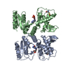
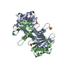
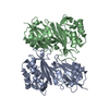
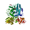
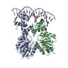
| Title | DEOXYNUCLEOSIDE MONOPHOSPHATE KINASE COMPLEXED WITH DEOXY-GMP AND AMP | ||||||
 Components Components | DEOXYNUCLEOSIDE MONOPHOSPHATE KINASE | ||||||
 Keywords Keywords |  PHOSPHOTRANSFERASE / TRANSFERASE PHOSPHOTRANSFERASE / TRANSFERASE | ||||||
| Function / homology |  Function and homology information(deoxy)nucleoside-phosphate kinase / (deoxy)nucleoside-phosphate kinase activity / phosphorylation / ATP binding Function and homology information(deoxy)nucleoside-phosphate kinase / (deoxy)nucleoside-phosphate kinase activity / phosphorylation / ATP bindingSimilarity search - Function | ||||||
| Biological species |  Enterobacteria phage T4 (virus) Enterobacteria phage T4 (virus) | ||||||
| Method | X-RAY DIFFRACTION / SYNCHROTRON / Resolution: 2.2 Å | ||||||
 Authors Authors | Teplyakov, A. / Sebastiao, P. | ||||||
 Citation Citation | Journal: EMBO J. / Year: 1996 Title: Crystal structure of bacteriophage T4 deoxynucleotide kinase with its substrates dGMP and ATP. Authors: Teplyakov, A. / Sebastiao, P. / Obmolova, G. / Perrakis, A. / Brush, G.S. / Bessman, M.J. / Wilson, K.S. #1: Journal: Acta Crystallogr.,Sect.D / Year: 1996Title: Crystallization and Preliminary X-Ray Analysis of Bacteriophage T4 Deoxynucleotide Kinase Authors: Sebastiao, P. / Obmolova, G. / Brush, G.S. / Bessman, M.J. / Teplyakov, A. | ||||||
| History |
|
- Structure visualization
| Structure viewer | Molecule: MolmilJmol/JSmol |
|---|
- Downloads & links
-Download
| PDBx/mmCIF format | 1del.cif.gz | 112.3 KB | Display | PDBx/mmCIF format |
|---|---|---|---|---|
| PDB format | pdb1del.ent.gz | 86.7 KB | Display | PDB format |
| PDBx/mmJSON format | 1del.json.gz | Tree view | PDBx/mmJSON format | |
| Others |  Other downloads Other downloads |
-Validation report
| Arichive directory | https://data.pdbj.org/pub/pdb/validation_reports/de/1delftp://data.pdbj.org/pub/pdb/validation_reports/de/1del | HTTPS FTP |
|---|
-Related structure data


-Links
 PDBj
PDBj


- Assembly
| Deposited unit | 
| ||||||||
|---|---|---|---|---|---|---|---|---|---|
| 1 |
| ||||||||
| Unit cell |
| ||||||||
| Noncrystallographic symmetry (NCS) | NCS oper: (Code: given Matrix: (-0.98807, -0.13116, 0.08074), Vector : |
-Components
| #1: Protein | Mass: 27359.111 Da / Num. of mol.: 2 Source method: isolated from a genetically manipulated source Source: (gene. exp.) Enterobacteria phage T4 (virus) / Genus: T4-like viruses / Species: Enterobacteria phage T4 sensu lato / Gene: 1 / Plasmid: PBK5 / Gene (production host): 1 / Production host:  Escherichia coli (E. coli) Escherichia coli (E. coli)References: UniProt: P04531, (deoxy)nucleoside-phosphate kinase#2: Chemical |   Mass: 24.305 Da / Num. of mol.: 2 / Source method: obtained synthetically / Formula: Mg Mass: 24.305 Da / Num. of mol.: 2 / Source method: obtained synthetically / Formula: Mg#3: Chemical | Deoxyguanosine monophosphate  Mass: 347.221 Da / Num. of mol.: 2 / Source method: obtained synthetically / Formula: C10H14N5O7P Mass: 347.221 Da / Num. of mol.: 2 / Source method: obtained synthetically / Formula: C10H14N5O7P#4: Chemical | ChemComp-AMP / | Adenosine monophosphate  Mass: 347.221 Da / Num. of mol.: 1 / Source method: obtained synthetically / Formula: C10H14N5O7P / Comment: AMP*YM Mass: 347.221 Da / Num. of mol.: 1 / Source method: obtained synthetically / Formula: C10H14N5O7P / Comment: AMP*YM#5: Water | ChemComp-HOH / | Water Mass: 18.015 Da / Num. of mol.: 222 / Source method: isolated from a natural source / Formula: H2O Mass: 18.015 Da / Num. of mol.: 222 / Source method: isolated from a natural source / Formula: H2O |
|---|
-Experimental details
-Experiment
| Experiment | Method: X-RAY DIFFRACTION |
|---|
- Sample preparation
| Crystal | Density Matthews: 2.98 Å3/Da / Density % sol: 59 % | ||||||||||||||||||||||||||||||||||||||||||||||||||
|---|---|---|---|---|---|---|---|---|---|---|---|---|---|---|---|---|---|---|---|---|---|---|---|---|---|---|---|---|---|---|---|---|---|---|---|---|---|---|---|---|---|---|---|---|---|---|---|---|---|---|---|
| Crystal grow | pH: 6.5 / Details: pH 6.5 | ||||||||||||||||||||||||||||||||||||||||||||||||||
| Crystal | *PLUS | ||||||||||||||||||||||||||||||||||||||||||||||||||
| Crystal grow | *PLUS Temperature: 4 ℃ / pH: 7.6 / Method: vapor diffusion, hanging drop | ||||||||||||||||||||||||||||||||||||||||||||||||||
| Components of the solutions | *PLUS
|
-Data collection
| Diffraction | Mean temperature: 277 K |
|---|---|
| Diffraction source | Source: SYNCHROTRON / Site: EMBL/DESY, HAMBURG  / Beamline: X11 / Wavelength: 0.92 / Beamline: X11 / Wavelength: 0.92 |
| Detector | Type: MAR scanner 180 mm plate / Detector: IMAGE PLATE / Date: Apr 1, 1995 / Details: TOROIDAL MIRROR |
| Radiation | Monochromator: SI(111) / Monochromatic (M) / Laue (L): M / Scattering type: x-ray |
| Radiation wavelength | Wavelength: 0.92 Å / Relative weight: 1 |
| Reflection | Resolution: 2.2→10 Å / Num. obs: 31836 / % possible obs: 96.5 % / Observed criterion σ(I): -3 / Redundancy: 3 % / Rmerge(I) obs: 0.065 |
| Reflection | *PLUS Num. measured all: 96619 |
- Processing
| Software |
| ||||||||||||||||||||||||||||||||||||||||||||||||||||||||||||||||||||||||||||||||||||
|---|---|---|---|---|---|---|---|---|---|---|---|---|---|---|---|---|---|---|---|---|---|---|---|---|---|---|---|---|---|---|---|---|---|---|---|---|---|---|---|---|---|---|---|---|---|---|---|---|---|---|---|---|---|---|---|---|---|---|---|---|---|---|---|---|---|---|---|---|---|---|---|---|---|---|---|---|---|---|---|---|---|---|---|---|---|
| Refinement | Resolution: 2.2→10 Å / σ(F): 0
| ||||||||||||||||||||||||||||||||||||||||||||||||||||||||||||||||||||||||||||||||||||
| Displacement parameters | Biso mean: 39.6 Å2 | ||||||||||||||||||||||||||||||||||||||||||||||||||||||||||||||||||||||||||||||||||||
| Refinement step | Cycle: LAST / Resolution: 2.2→10 Å
| ||||||||||||||||||||||||||||||||||||||||||||||||||||||||||||||||||||||||||||||||||||
| Refine LS restraints |
| ||||||||||||||||||||||||||||||||||||||||||||||||||||||||||||||||||||||||||||||||||||
| Software | *PLUS Name: PROLSQ / Classification: refinement | ||||||||||||||||||||||||||||||||||||||||||||||||||||||||||||||||||||||||||||||||||||
| Refinement | *PLUS σ(F): 3 / Rfactor all: 0.186 / Rfactor obs: 0.173 | ||||||||||||||||||||||||||||||||||||||||||||||||||||||||||||||||||||||||||||||||||||
| Solvent computation | *PLUS | ||||||||||||||||||||||||||||||||||||||||||||||||||||||||||||||||||||||||||||||||||||
| Displacement parameters | *PLUS | ||||||||||||||||||||||||||||||||||||||||||||||||||||||||||||||||||||||||||||||||||||
| Refine LS restraints | *PLUS Type: p_angle_d / Dev ideal: 0.027 |
