 Movie
Movie Controller

[English] 日本語
 Yorodumi
- PDB-1dc9: PROPERTIES AND CRYSTAL STRUCTURE OF A BETA-BARREL FOLDING MUTANT,... -
+ Open data

- Basic information
| Entry | Database: PDB / ID: 1dc9 | ||||||
|---|---|---|---|---|---|---|---|
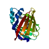
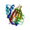
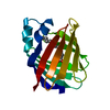
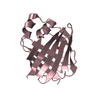
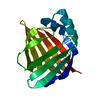
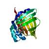
| Title | PROPERTIES AND CRYSTAL STRUCTURE OF A BETA-BARREL FOLDING MUTANT, V60N INTESTINAL FATTY ACID BINDING PROTEIN (IFABP) | ||||||
 Components Components | INTESTINAL FATTY ACID BINDING PROTEIN | ||||||
 Keywords Keywords | LIPID BINDING PROTEIN /  FATTY ACID BINDING PROTEIN / INTRACELLULAR LIPID BINDING PROTEIN / MUTANT / BETA- BARREL FATTY ACID BINDING PROTEIN / INTRACELLULAR LIPID BINDING PROTEIN / MUTANT / BETA- BARREL | ||||||
| Function / homology |  Function and homology information Function and homology informationTriglyceride catabolism / intestinal lipid absorption / apical cortex / intestinal absorption / long-chain fatty acid transmembrane transporter activity / long-chain fatty acid binding / microvillus / fatty acid transport / long-chain fatty acid transport / fatty acid metabolic process ...Triglyceride catabolism / intestinal lipid absorption / apical cortex / intestinal absorption / long-chain fatty acid transmembrane transporter activity / long-chain fatty acid binding / microvillus / fatty acid transport / long-chain fatty acid transport / fatty acid metabolic process / fatty acid binding / nucleus / cytosolSimilarity search - Function | ||||||
| Biological species |  Rattus norvegicus (Norway rat) Rattus norvegicus (Norway rat) | ||||||
| Method | X-RAY DIFFRACTION / Resolution: 2.1 Å | ||||||
 Authors Authors | Ropson, I.J. / Yowler, B.C. / Dalessio, P.M. / Banaszak, L. / Thompson, J. | ||||||
 Citation Citation | Journal: Biophys.J. / Year: 2000 Title: Properties and crystal structure of a beta-barrel folding mutant. Authors: Ropson, I.J. / Yowler, B.C. / Dalessio, P.M. / Banaszak, L. / Thompson, J. | ||||||
| History |
|
- Structure visualization
| Structure viewer | Molecule: MolmilJmol/JSmol |
|---|
- Downloads & links
-Download
| PDBx/mmCIF format | 1dc9.cif.gz | 38.8 KB | Display | PDBx/mmCIF format |
|---|---|---|---|---|
| PDB format | pdb1dc9.ent.gz | 26.7 KB | Display | PDB format |
| PDBx/mmJSON format | 1dc9.json.gz | Tree view | PDBx/mmJSON format | |
| Others |  Other downloads Other downloads |
-Validation report
| Arichive directory | https://data.pdbj.org/pub/pdb/validation_reports/dc/1dc9ftp://data.pdbj.org/pub/pdb/validation_reports/dc/1dc9 | HTTPS FTP |
|---|
-Related structure data
| Related structure data | |
|---|---|
| Similar structure data |


-Links
 PDBj
PDBj

- Assembly
| Deposited unit | 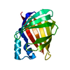
| ||||||||
|---|---|---|---|---|---|---|---|---|---|
| 1 |
| ||||||||
| Unit cell |
|
-Components
| #1: Protein | Mass: 15029.987 Da / Num. of mol.: 1 / Mutation: V60N Source method: isolated from a genetically manipulated source Source: (gene. exp.) Rattus norvegicus (Norway rat) / Organ: INTESTINE / Production host:  Escherichia coli (E. coli) / References: UniProt: P02693 Escherichia coli (E. coli) / References: UniProt: P02693 |
|---|---|
| #2: Water | ChemComp-HOH / Water Mass: 18.015 Da / Num. of mol.: 73 / Source method: isolated from a natural source / Formula: H2O Mass: 18.015 Da / Num. of mol.: 73 / Source method: isolated from a natural source / Formula: H2O |
-Experimental details
-Experiment
| Experiment | Method: X-RAY DIFFRACTION / Number of used crystals: 2 |
|---|
- Sample preparation
| Crystal | Density Matthews: 2.02 Å3/Da / Density % sol: 39 % | ||||||||||||||||||||||||
|---|---|---|---|---|---|---|---|---|---|---|---|---|---|---|---|---|---|---|---|---|---|---|---|---|---|
| Crystal grow | Method: vapor diffusion, hanging drop / pH: 7.3 Details: 36-38% PEG 4000, 0.1 M PIPES, 1:1 RATIO WELL AND 4.6 MG/ML PROTEIN IN WATER, pH 7.3, VAPOR DIFFUSION, HANGING DROP | ||||||||||||||||||||||||
| Crystal grow | *PLUS | ||||||||||||||||||||||||
| Components of the solutions | *PLUS
|
-Data collection
| Diffraction | Mean temperature: 298 K |
|---|---|
| Diffraction source | Source: ROTATING ANODE / Type: RIGAKU / Wavelength: 1.5418 |
| Detector | Type: SIEMENS HI-STAR / Detector: AREA DETECTOR / Date: Mar 5, 1999 |
| Radiation | Protocol: SINGLE WAVELENGTH / Monochromatic (M) / Laue (L): M / Scattering type: x-ray |
| Radiation wavelength | Wavelength: 1.5418 Å / Relative weight: 1 |
| Reflection | Resolution: 2.1→20 Å / Num. all: 6804 / Num. obs: 6804 / % possible obs: 91 % / Observed criterion σ(F): 0 / Observed criterion σ(I): 0 / Redundancy: 3.1 % / Biso Wilson estimate: 5.7 Å2 / Rmerge(I) obs: 0.034 / Net I/σ(I): 23.9 |
| Reflection shell | Resolution: 2.09→2.22 Å / Redundancy: 1.7 % / Rmerge(I) obs: 0.145 / % possible all: 38.2 |
| Reflection | *PLUS Num. measured all: 19796 / Rmerge(I) obs: 0.047 |
| Reflection shell | *PLUS % possible obs: 38.2 % / Num. unique obs: 447 / Num. measured obs: 779 / Rmerge(I) obs: 0.151 / Mean I/σ(I) obs: 1.4 |
- Processing
| Software |
| ||||||||||||||||||||||||||||||||||||||||||||||||||||||||||||||||||||||||||||||||
|---|---|---|---|---|---|---|---|---|---|---|---|---|---|---|---|---|---|---|---|---|---|---|---|---|---|---|---|---|---|---|---|---|---|---|---|---|---|---|---|---|---|---|---|---|---|---|---|---|---|---|---|---|---|---|---|---|---|---|---|---|---|---|---|---|---|---|---|---|---|---|---|---|---|---|---|---|---|---|---|---|---|
| Refinement | Resolution: 2.1→20 Å / Rfactor Rfree error: 0.01 / Data cutoff high absF: 158121.65 / Data cutoff high rms absF: 158121.65 / Data cutoff low absF: 0 / Isotropic thermal model: RESTRAINED / Cross valid method: THROUGHOUT / σ(F): 0 / σ(I): 0 / Stereochemistry target values: ENGH & HUBER
| ||||||||||||||||||||||||||||||||||||||||||||||||||||||||||||||||||||||||||||||||
| Displacement parameters | Biso mean: 18.5 Å2
| ||||||||||||||||||||||||||||||||||||||||||||||||||||||||||||||||||||||||||||||||
| Refine analyze |
| ||||||||||||||||||||||||||||||||||||||||||||||||||||||||||||||||||||||||||||||||
| Refinement step | Cycle: LAST / Resolution: 2.1→20 Å
| ||||||||||||||||||||||||||||||||||||||||||||||||||||||||||||||||||||||||||||||||
| Refine LS restraints |
| ||||||||||||||||||||||||||||||||||||||||||||||||||||||||||||||||||||||||||||||||
| LS refinement shell | Resolution: 2.1→2.23 Å / Rfactor Rfree error: 0.049 / Total num. of bins used: 6
| ||||||||||||||||||||||||||||||||||||||||||||||||||||||||||||||||||||||||||||||||
| Xplor file |
| ||||||||||||||||||||||||||||||||||||||||||||||||||||||||||||||||||||||||||||||||
| Software | *PLUS Name: CNS / Version: 0.3 / Classification: refinement | ||||||||||||||||||||||||||||||||||||||||||||||||||||||||||||||||||||||||||||||||
| Refinement | *PLUS σ(F): 0 / % reflection Rfree: 8 % | ||||||||||||||||||||||||||||||||||||||||||||||||||||||||||||||||||||||||||||||||
| Solvent computation | *PLUS | ||||||||||||||||||||||||||||||||||||||||||||||||||||||||||||||||||||||||||||||||
| Displacement parameters | *PLUS Biso mean: 18.5 Å2 | ||||||||||||||||||||||||||||||||||||||||||||||||||||||||||||||||||||||||||||||||
| Refine LS restraints | *PLUS
| ||||||||||||||||||||||||||||||||||||||||||||||||||||||||||||||||||||||||||||||||
| LS refinement shell | *PLUS Rfactor Rfree: 0.362 / % reflection Rfree: 10.6 % / Rfactor Rwork: 0.251 |
