 Movie
Movie Controller

[English] 日本語
 Yorodumi
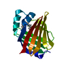
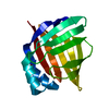
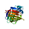
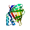
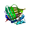
- PDB-1alb: CRYSTAL STRUCTURE OF RECOMBINANT MURINE ADIPOCYTE LIPID-BINDING P... -
+ Open data

- Basic information
| Entry | Database: PDB / ID: 1alb | ||||||
|---|---|---|---|---|---|---|---|
| Title | CRYSTAL STRUCTURE OF RECOMBINANT MURINE ADIPOCYTE LIPID-BINDING PROTEIN | ||||||
 Components Components | ADIPOCYTE LIPID-BINDING PROTEIN | ||||||
 Keywords Keywords | LIPID BINDING PROTEIN / LIPID-BINDING PROTEIN | ||||||
| Function / homology |  Function and homology information Function and homology informationTriglyceride catabolism /  hormone receptor binding / long-chain fatty acid transmembrane transporter activity / long-chain fatty acid binding / cellular response to lithium ion / white fat cell differentiation / fatty acid transport / long-chain fatty acid transport / brown fat cell differentiation / fatty acid metabolic process ...Triglyceride catabolism / hormone receptor binding / long-chain fatty acid transmembrane transporter activity / long-chain fatty acid binding / cellular response to lithium ion / white fat cell differentiation / fatty acid transport / long-chain fatty acid transport / brown fat cell differentiation / fatty acid metabolic process / cholesterol homeostasis / fatty acid binding / response to bacterium / positive regulation of inflammatory response / positive regulation of cold-induced thermogenesis / cellular response to tumor necrosis factor / negative regulation of DNA-templated transcription / positive regulation of cell population proliferation / nucleoplasm / nucleus / cytosol / cytoplasm hormone receptor binding / long-chain fatty acid transmembrane transporter activity / long-chain fatty acid binding / cellular response to lithium ion / white fat cell differentiation / fatty acid transport / long-chain fatty acid transport / brown fat cell differentiation / fatty acid metabolic process ...Triglyceride catabolism / hormone receptor binding / long-chain fatty acid transmembrane transporter activity / long-chain fatty acid binding / cellular response to lithium ion / white fat cell differentiation / fatty acid transport / long-chain fatty acid transport / brown fat cell differentiation / fatty acid metabolic process / cholesterol homeostasis / fatty acid binding / response to bacterium / positive regulation of inflammatory response / positive regulation of cold-induced thermogenesis / cellular response to tumor necrosis factor / negative regulation of DNA-templated transcription / positive regulation of cell population proliferation / nucleoplasm / nucleus / cytosol / cytoplasmSimilarity search - Function | ||||||
| Biological species |  Mus musculus (house mouse) Mus musculus (house mouse) | ||||||
| Method | X-RAY DIFFRACTION / Resolution: 2.5 Å | ||||||
 Authors Authors | Xu, Z. / Banaszak, L.J. | ||||||
 Citation Citation | Journal: Biochemistry / Year: 1992 Title: Crystal structure of recombinant murine adipocyte lipid-binding protein. Authors: Xu, Z. / Bernlohr, D.A. / Banaszak, L.J. #1: Journal: J.Biol.Chem. / Year: 1991Title: Expression, Purification, and Crystallization of the Adipocyte Lipid Binding Protein Authors: Xu, Z. / Buelt, M.K. / Banaszak, L.J. / Bernlohr, D.A. | ||||||
| History |
|
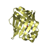
- Structure visualization
| Structure viewer | Molecule: MolmilJmol/JSmol |
|---|
- Downloads & links
-Download
| PDBx/mmCIF format | 1alb.cif.gz | 37.4 KB | Display | PDBx/mmCIF format |
|---|---|---|---|---|
| PDB format | pdb1alb.ent.gz | 26.2 KB | Display | PDB format |
| PDBx/mmJSON format | 1alb.json.gz | Tree view | PDBx/mmJSON format | |
| Others |  Other downloads Other downloads |
-Validation report
| Arichive directory | https://data.pdbj.org/pub/pdb/validation_reports/al/1albftp://data.pdbj.org/pub/pdb/validation_reports/al/1alb | HTTPS FTP |
|---|
-Related structure data
| Similar structure data |
|---|


-Links
 PDBj
PDBj

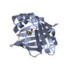
- Assembly
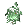
| Deposited unit | 
| ||||||||
|---|---|---|---|---|---|---|---|---|---|
| 1 |
| ||||||||
| Unit cell |
| ||||||||
| Atom site foot note | 1: THE SULFHYDRYL GROUP OF CYS 1 IS CHEMICALLY MODIFIED BUT NOT SHOWN IN THE ELECTRON DENSITY MAP. |
-Components
| #1: Protein | Mass: 14539.688 Da / Num. of mol.: 1 Source method: isolated from a genetically manipulated source Source: (gene. exp.) Mus musculus (house mouse) / Production host:  Escherichia coli (E. coli) / References: UniProt: P04117 Escherichia coli (E. coli) / References: UniProt: P04117 |
|---|---|
| #2: Water | ChemComp-HOH / Water Mass: 18.015 Da / Num. of mol.: 69 / Source method: isolated from a natural source / Formula: H2O Mass: 18.015 Da / Num. of mol.: 69 / Source method: isolated from a natural source / Formula: H2O |
| Sequence details | THE SULFHYDRYL |
-Experimental details
-Experiment
| Experiment | Method: X-RAY DIFFRACTION |
|---|
- Sample preparation
| Crystal | Density Matthews: 2.47 Å3/Da / Density % sol: 50.26 % | |||||||||||||||||||||||||||||||||||||||||||||
|---|---|---|---|---|---|---|---|---|---|---|---|---|---|---|---|---|---|---|---|---|---|---|---|---|---|---|---|---|---|---|---|---|---|---|---|---|---|---|---|---|---|---|---|---|---|---|
| Crystal grow | *PLUS Temperature: 19 ℃ / pH: 7 / Method: vapor diffusion, hanging drop / Details: used as seeds | |||||||||||||||||||||||||||||||||||||||||||||
| Components of the solutions | *PLUS
|
-Data collection
| Radiation | Scattering type: x-ray |
|---|---|
| Radiation wavelength | Relative weight: 1 |
| Reflection | *PLUS Highest resolution: 2.5 Å / Lowest resolution: 9999 Å / Num. all: 5227 / Num. measured all: 5115 |
- Processing
| Software |
| ||||||||||||||||||||||||||||||||||||||||||||||||||||||||||||
|---|---|---|---|---|---|---|---|---|---|---|---|---|---|---|---|---|---|---|---|---|---|---|---|---|---|---|---|---|---|---|---|---|---|---|---|---|---|---|---|---|---|---|---|---|---|---|---|---|---|---|---|---|---|---|---|---|---|---|---|---|---|
| Refinement | Resolution: 2.5→8 Å / Rfactor Rwork: 0.183 / Rfactor obs: 0.183 / σ(F): 0 | ||||||||||||||||||||||||||||||||||||||||||||||||||||||||||||
| Refinement step | Cycle: LAST / Resolution: 2.5→8 Å
| ||||||||||||||||||||||||||||||||||||||||||||||||||||||||||||
| Refine LS restraints |
| ||||||||||||||||||||||||||||||||||||||||||||||||||||||||||||
| Refinement | *PLUS Highest resolution: 2.5 Å / Lowest resolution: 8 Å / Num. reflection all: 4773 / σ(F): 0 / Rfactor all: 0.183 | ||||||||||||||||||||||||||||||||||||||||||||||||||||||||||||
| Solvent computation | *PLUS | ||||||||||||||||||||||||||||||||||||||||||||||||||||||||||||
| Displacement parameters | *PLUS | ||||||||||||||||||||||||||||||||||||||||||||||||||||||||||||
| Refine LS restraints | *PLUS
|
