 Movie
Movie Controller


+ Open data

- Basic information
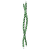
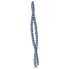
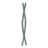
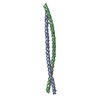
| Entry | Database: PDB / ID: 1d7m | ||||||
|---|---|---|---|---|---|---|---|
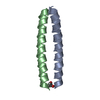
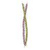
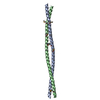
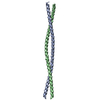
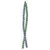
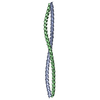
| Title | COILED-COIL DIMERIZATION DOMAIN FROM CORTEXILLIN I | ||||||
 Components Components | CORTEXILLIN I | ||||||
 Keywords Keywords |  CONTRACTILE PROTEIN / COILED-COIL / COILED-COIL TRIGGER SITE / ALPHA HELIX / DIMERIZATION CONTRACTILE PROTEIN / COILED-COIL / COILED-COIL TRIGGER SITE / ALPHA HELIX / DIMERIZATION | ||||||
| Function / homology |  Function and homology information Function and homology informationlateral part of motile cell / sorocarp stalk morphogenesis / negative regulation of protein localization to cell cortex / protein localization to cleavage furrow / actomyosin contractile ring contraction / adenylate cyclase-activating G protein-coupled cAMP receptor signaling pathway / regulation of myosin II filament assembly / chemotaxis to cAMP / positive regulation of cytoskeleton organization / protein localization => GO:0008104 ...lateral part of motile cell / sorocarp stalk morphogenesis / negative regulation of protein localization to cell cortex / protein localization to cleavage furrow / actomyosin contractile ring contraction / adenylate cyclase-activating G protein-coupled cAMP receptor signaling pathway / regulation of myosin II filament assembly / chemotaxis to cAMP / positive regulation of cytoskeleton organization / protein localization => GO:0008104 / cell trailing edge / actin filament network formation / basal cortex / aggregation involved in sorocarp development / actomyosin contractile ring / mitotic spindle midzone / sexual reproduction / actin crosslink formation / detection of mechanical stimulus / cleavage furrow formation / bleb assembly / alpha-catenin binding / epithelial cell morphogenesis / protein kinase regulator activity / cortical actin cytoskeleton organization / regulation of GTPase activity / cell leading edge / actin filament bundle / cleavage furrow / actin filament bundle assembly / mitotic cytokinesis / response to mechanical stimulus / phagocytic vesicle / phosphatidylinositol-4,5-bisphosphate binding / actin filament / response to bacterium / cell morphogenesis / establishment of protein localization / protein localization / actin filament binding / actin cytoskeleton / cell cortex / regulation of gene expression / vesicle / positive regulation of protein phosphorylation / positive regulation of gene expression / identical protein binding / plasma membrane / cytosol / cytoplasmSimilarity search - Function | ||||||
| Biological species |  Dictyostelium discoideum (eukaryote) Dictyostelium discoideum (eukaryote) | ||||||
| Method | X-RAY DIFFRACTION / SYNCHROTRON / Resolution: 2.7 Å | ||||||
 Authors Authors | Burkhard, P. / Kammerer, R.A. / Steinmetz, M.O. / Bourenkov, G.P. / Aebi, U. | ||||||
 Citation Citation | Journal: Structure Fold.Des. / Year: 2000 Title: The coiled-coil trigger site of the rod domain of cortexillin I unveils a distinct network of interhelical and intrahelical salt bridges. Authors: Burkhard, P. / Kammerer, R.A. / Steinmetz, M.O. / Bourenkov, G.P. / Aebi, U. #1: Journal: J.Struct.Biol. / Year: 1998Title: Crystallization and Preliminary X-Ray Diffraction Analysis of the 190 A Long Coiled-Coil Dimerization Domain of the Actin-Bundling Protein Cortexillin I from Dictyostelium discoideum Authors: Burkhard, P. / Steinmetz, M.O. / Schulthess, T. / Landwehr, R. / Aebi, U. / Kammerer, R.A. | ||||||
| History |
|
- Structure visualization
| Structure viewer | Molecule: MolmilJmol/JSmol |
|---|
- Downloads & links
-Download
| PDBx/mmCIF format | 1d7m.cif.gz | 51.7 KB | Display | PDBx/mmCIF format |
|---|---|---|---|---|
| PDB format | pdb1d7m.ent.gz | 39.5 KB | Display | PDB format |
| PDBx/mmJSON format | 1d7m.json.gz | Tree view | PDBx/mmJSON format | |
| Others |  Other downloads Other downloads |
-Validation report
| Arichive directory | https://data.pdbj.org/pub/pdb/validation_reports/d7/1d7mftp://data.pdbj.org/pub/pdb/validation_reports/d7/1d7m | HTTPS FTP |
|---|
-Related structure data
| Related structure data | |
|---|---|
| Similar structure data |

-Links
 PDBj
PDBj- Assembly
| Deposited unit | 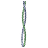
| ||||||||
|---|---|---|---|---|---|---|---|---|---|
| 1 |
| ||||||||
| 2 | 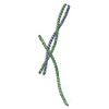
| ||||||||
| Unit cell |
| ||||||||
| Components on special symmetry positions |
|
-Components
| #1: Protein | Mass: 11623.278 Da / Num. of mol.: 2 / Fragment: COILED-COIL DIMERIZATION DOMAIN Source method: isolated from a genetically manipulated source Source: (gene. exp.) Dictyostelium discoideum (eukaryote) / Production host:  Escherichia coli (E. coli) / References: UniProt: O15813, UniProt: Q54HG2*PLUS Escherichia coli (E. coli) / References: UniProt: O15813, UniProt: Q54HG2*PLUS#2: Water | ChemComp-HOH / | Water Mass: 18.015 Da / Num. of mol.: 93 / Source method: isolated from a natural source / Formula: H2O Mass: 18.015 Da / Num. of mol.: 93 / Source method: isolated from a natural source / Formula: H2O |
|---|
-Experimental details
-Experiment
| Experiment | Method: X-RAY DIFFRACTION / Number of used crystals: 2 |
|---|
- Sample preparation
| Crystal | Density Matthews: 4.46 Å3/Da / Density % sol: 72.43 % | ||||||||||||||||||||||||||||||||||||||||||||||||
|---|---|---|---|---|---|---|---|---|---|---|---|---|---|---|---|---|---|---|---|---|---|---|---|---|---|---|---|---|---|---|---|---|---|---|---|---|---|---|---|---|---|---|---|---|---|---|---|---|---|
| Crystal grow | Temperature: 297 K / Method: vapor diffusion, hanging drop / pH: 6.5 Details: 1.5 M AMMONIUM SULFATE 100 MM TRIS (PH 6.5) 2.5 % PEG 400 1.0 % DIOXANE, VAPOR DIFFUSION, HANGING DROP, temperature 297K | ||||||||||||||||||||||||||||||||||||||||||||||||
| Crystal grow | *PLUS pH: 7.4 / Details: Burkhard, P., (1998) J.Struct.Biol., 122, 293. | ||||||||||||||||||||||||||||||||||||||||||||||||
| Components of the solutions | *PLUS
|
-Data collection
| Diffraction |
| |||||||||||||||
|---|---|---|---|---|---|---|---|---|---|---|---|---|---|---|---|---|
| Diffraction source |
| |||||||||||||||
| Detector |
| |||||||||||||||
| Radiation |
| |||||||||||||||
| Radiation wavelength |
| |||||||||||||||
| Reflection | Resolution: 2.7→30 Å / Num. all: 31909 / Num. obs: 11302 / % possible obs: 95.9 % / Redundancy: 2.8 % / Biso Wilson estimate: 37.9 Å2 / Rmerge(I) obs: 0.111 / Net I/σ(I): 5.3 | |||||||||||||||
| Reflection shell | Resolution: 2.7→2.85 Å / Redundancy: 2.7 % / Rmerge(I) obs: 0.462 / % possible all: 88.2 | |||||||||||||||
| Reflection | *PLUS % possible obs: 94.6 % / Num. measured all: 49645 / Rmerge(I) obs: 0.078 |

- Processing
| Software |
| ||||||||||||||||||||||||||||||||||||||||||||||||||||||||||||||||||||||||||||||||
|---|---|---|---|---|---|---|---|---|---|---|---|---|---|---|---|---|---|---|---|---|---|---|---|---|---|---|---|---|---|---|---|---|---|---|---|---|---|---|---|---|---|---|---|---|---|---|---|---|---|---|---|---|---|---|---|---|---|---|---|---|---|---|---|---|---|---|---|---|---|---|---|---|---|---|---|---|---|---|---|---|---|
| Refinement | Resolution: 2.7→37.3 Å / Rfactor Rfree error: 0.011 / Data cutoff high absF: 1675937.19 / Data cutoff high rms absF: 1675937.19 / Data cutoff low absF: 0 / Isotropic thermal model: CONSTRAINED / Cross valid method: THROUGHOUT / σ(F): 3
| ||||||||||||||||||||||||||||||||||||||||||||||||||||||||||||||||||||||||||||||||
| Solvent computation | Solvent model: FLAT MODEL / Bsol: 71.69 Å2 / ksol: 0.406 e/Å3 | ||||||||||||||||||||||||||||||||||||||||||||||||||||||||||||||||||||||||||||||||
| Displacement parameters | Biso mean: 56.9 Å2
| ||||||||||||||||||||||||||||||||||||||||||||||||||||||||||||||||||||||||||||||||
| Refine analyze |
| ||||||||||||||||||||||||||||||||||||||||||||||||||||||||||||||||||||||||||||||||
| Refinement step | Cycle: LAST / Resolution: 2.7→37.3 Å
| ||||||||||||||||||||||||||||||||||||||||||||||||||||||||||||||||||||||||||||||||
| Refine LS restraints |
| ||||||||||||||||||||||||||||||||||||||||||||||||||||||||||||||||||||||||||||||||
| Refine LS restraints NCS | NCS model details: CONSTRAINED | ||||||||||||||||||||||||||||||||||||||||||||||||||||||||||||||||||||||||||||||||
| LS refinement shell | Resolution: 2.7→2.87 Å / Rfactor Rfree error: 0.052 / Total num. of bins used: 6
| ||||||||||||||||||||||||||||||||||||||||||||||||||||||||||||||||||||||||||||||||
| Xplor file |
| ||||||||||||||||||||||||||||||||||||||||||||||||||||||||||||||||||||||||||||||||
| Software | *PLUS Name: CNS / Version: 0.9 / Classification: refinement | ||||||||||||||||||||||||||||||||||||||||||||||||||||||||||||||||||||||||||||||||
| Refinement | *PLUS Lowest resolution: 30 Å / σ(F): 3 / % reflection Rfree: 5 % / Rfactor obs: 0.218 / Rfactor Rfree: 0.257 | ||||||||||||||||||||||||||||||||||||||||||||||||||||||||||||||||||||||||||||||||
| Solvent computation | *PLUS | ||||||||||||||||||||||||||||||||||||||||||||||||||||||||||||||||||||||||||||||||
| Displacement parameters | *PLUS Biso mean: 56.9 Å2 | ||||||||||||||||||||||||||||||||||||||||||||||||||||||||||||||||||||||||||||||||
| Refine LS restraints | *PLUS
| ||||||||||||||||||||||||||||||||||||||||||||||||||||||||||||||||||||||||||||||||
| LS refinement shell | *PLUS Rfactor Rfree: 0.381 / % reflection Rfree: 5.1 % / Rfactor Rwork: 0.268 |
