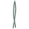
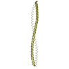
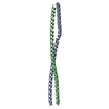
- PDB-3q8t: Crystal structure of the coiled coil domain of Beclin 1, an essen... -
+
Open data
ID or keywords:
Loading...
-
Basic information
Entry
Database: PDB / ID: 3q8t
Title
Crystal structure of the coiled coil domain of Beclin 1, an essential autophagy protein
Components
Beclin-1BECN1
Keywords
APOPTOSIS / Autophagy / Atg14L UVRAG
Function / homology
Function and homology information
cellular response to aluminum ion / Macroautophagy / : / phosphatidylinositol 3-kinase complex, class III / phosphatidylinositol 3-kinase complex, class III, type II / phosphatidylinositol 3-kinase complex, class III, type I / : / response to mitochondrial depolarisation / positive regulation of attachment of mitotic spindle microtubules to kinetochore / Ub-specific processing proteases ...cellular response to aluminum ion / Macroautophagy / : / phosphatidylinositol 3-kinase complex, class III / phosphatidylinositol 3-kinase complex, class III, type II / phosphatidylinositol 3-kinase complex, class III, type I / : / response to mitochondrial depolarisation / positive regulation of attachment of mitotic spindle microtubules to kinetochore / Ub-specific processing proteases / response to xenobiotic stimulus => GO:0009410 / negative regulation of lysosome organization / positive regulation of autophagosome assembly / engulfment of apoptotic cell / negative regulation of autophagosome assembly / receptor catabolic process / autophagy of mitochondrion / early endosome to late endosome transport / cellular response to nitrogen starvation / late endosome to vacuole transport / response to other organism / regulation of catalytic activity / phagophore assembly site / response to iron(II) ion / mitotic metaphase chromosome alignment / lysosome organization / extrinsic component of membrane / positive regulation of cardiac muscle hypertrophy / mitophagy / autophagosome assembly / autophagosome / neuron development / negative regulation of reactive oxygen species metabolic process / response to vitamin E / cellular response to glucose starvation / amyloid-beta metabolic process / phosphatidylinositol 3-kinase binding / phagocytic vesicle / positive regulation of autophagy / positive regulation of intrinsic apoptotic signaling pathway / cellular response to epidermal growth factor stimulus / cellular response to copper ion / cellular response to amino acid starvation / response to nutrient levels / negative regulation of autophagy / regulation of cytokinesis / mitochondrial membrane / macroautophagy / response to lead ion / trans-Golgi network / cellular response to hydrogen peroxide / autophagy / GTPase binding / cytoplasmic vesicle / defense response to virus / response to hypoxia / endosome membrane / endosome / cell division / negative regulation of cell population proliferation / dendrite / apoptotic process / ubiquitin protein ligase binding / endoplasmic reticulum membrane / negative regulation of apoptotic process / protein kinase binding / endoplasmic reticulum / protein-containing complex / identical protein binding / nucleus / cytosol / cytoplasm Similarity search - Function
Beclin-1 / Single alpha-helices involved in coiled-coils or other helix-helix interfaces - #3110 / Beclin-1, BH3 domain / Beclin-1 BH3 domain, Bcl-2-interacting / Atg6/Beclin / Atg6/Beclin C-terminal domain superfamily / Atg6, BARA domain / Atg6/beclin, coiled-coil domain / Apg6 BARA domain / Apg6 coiled-coil region ...Beclin-1 / Single alpha-helices involved in coiled-coils or other helix-helix interfaces - #3110 / Beclin-1, BH3 domain / Beclin-1 BH3 domain, Bcl-2-interacting / Atg6/Beclin / Atg6/Beclin C-terminal domain superfamily / Atg6, BARA domain / Atg6/beclin, coiled-coil domain / Apg6 BARA domain / Apg6 coiled-coil region / Single alpha-helices involved in coiled-coils or other helix-helix interfaces / Helix non-globular / Special Similarity search - Domain/homology
Method to determine structure: MIR / Resolution: 1.9→35.92 Å / Cor.coef. Fo:Fc: 0.941 / Cor.coef. Fo:Fc free: 0.933 / SU B: 2.666 / SU ML: 0.083 / Cross valid method: THROUGHOUT / ESU R Free: 0.142 / Stereochemistry target values: MAXIMUM LIKELIHOOD / Details: HYDROGENS HAVE BEEN ADDED IN THE RIDING POSITIONS
Rfactor
Num. reflection
% reflection
Selection details
Rfree
0.23631
1062
5.2 %
RANDOM
Rwork
0.20876
-
-
-
obs
0.21024
19438
100 %
-
Solvent computation
Ion probe radii: 0.8 Å / Shrinkage radii: 0.8 Å / VDW probe radii: 1.2 Å / Solvent model: MASK
Displacement parameters
Biso mean: 25.905 Å2
Baniso -1
Baniso -2
Baniso -3
1-
-0.01 Å2
0 Å2
0.05 Å2
2-
-
0.01 Å2
0 Å2
3-
-
-
0.02 Å2
Refinement step
Cycle: LAST / Resolution: 1.9→35.92 Å
Protein
Nucleic acid
Ligand
Solvent
Total
Num. atoms
1592
0
0
279
1871
Refine LS restraints
Refine-ID
Type
Dev ideal
Dev ideal target
Number
X-RAY DIFFRACTION
r_bond_refined_d
0.014
0.022
1599
X-RAY DIFFRACTION
r_angle_refined_deg
1.243
1.996
2128
X-RAY DIFFRACTION
r_dihedral_angle_1_deg
3.627
5
187
X-RAY DIFFRACTION
r_dihedral_angle_2_deg
37.46
26.538
104
X-RAY DIFFRACTION
r_dihedral_angle_3_deg
18.011
15
358
X-RAY DIFFRACTION
r_dihedral_angle_4_deg
19.803
15
14
X-RAY DIFFRACTION
r_chiral_restr
0.091
0.2
224
X-RAY DIFFRACTION
r_gen_planes_refined
0.005
0.02
1212
X-RAY DIFFRACTION
r_nbd_refined
0.243
0.2
871
X-RAY DIFFRACTION
r_nbtor_refined
0.291
0.2
1130
X-RAY DIFFRACTION
r_xyhbond_nbd_refined
0.199
0.2
218
X-RAY DIFFRACTION
r_symmetry_vdw_refined
0.175
0.2
50
X-RAY DIFFRACTION
r_symmetry_hbond_refined
0.194
0.2
32
X-RAY DIFFRACTION
r_mcbond_it
1.243
1.5
987
X-RAY DIFFRACTION
r_mcangle_it
1.732
2
1500
X-RAY DIFFRACTION
r_scbond_it
3.529
3
678
X-RAY DIFFRACTION
r_scangle_it
5.434
4.5
628
LS refinement shell
Resolution: 1.9→1.949 Å / Total num. of bins used: 20
Rfactor
Num. reflection
% reflection
Rfree
0.262
88
-
Rwork
0.207
1384
-
obs
-
-
100 %
+
About Yorodumi
-
News
-
Feb 9, 2022. New format data for meta-information of EMDB entries
New format data for meta-information of EMDB entries
Version 3 of the EMDB header file is now the official format.
The previous official version 1.9 will be removed from the archive.
In the structure databanks used in Yorodumi, some data are registered as the other names, "COVID-19 virus" and "2019-nCoV". Here are the details of the virus and the list of structure data.
Jan 31, 2019. EMDB accession codes are about to change! (news from PDBe EMDB page)
EMDB accession codes are about to change! (news from PDBe EMDB page)
The allocation of 4 digits for EMDB accession codes will soon come to an end. Whilst these codes will remain in use, new EMDB accession codes will include an additional digit and will expand incrementally as the available range of codes is exhausted. The current 4-digit format prefixed with “EMD-” (i.e. EMD-XXXX) will advance to a 5-digit format (i.e. EMD-XXXXX), and so on. It is currently estimated that the 4-digit codes will be depleted around Spring 2019, at which point the 5-digit format will come into force.
The EM Navigator/Yorodumi systems omit the EMD- prefix.
Related info.:Q: What is EMD? / ID/Accession-code notation in Yorodumi/EM Navigator
Yorodumi is a browser for structure data from EMDB, PDB, SASBDB, etc.
This page is also the successor to EM Navigator detail page, and also detail information page/front-end page for Omokage search.
The word "yorodu" (or yorozu) is an old Japanese word meaning "ten thousand". "mi" (miru) is to see.
Related info.:EMDB / PDB / SASBDB / Comparison of 3 databanks / Yorodumi Search / Aug 31, 2016. New EM Navigator & Yorodumi / Yorodumi Papers / Jmol/JSmol / Function and homology information / Changes in new EM Navigator and Yorodumi
 Movie
Movie Controller

 Yorodumi
 Open data
 Basic information
 Components
 BECN1
BECN1  Keywords
 Function and homology information
 Authors
 Citation
 Structure visualization
 Downloads & links
 Other downloads
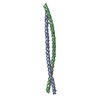

 PDBj
PDBj

 Assembly

 Mass: 18.015 Da / Num. of mol.: 279 / Source method: isolated from a natural source / Formula: H2O
 Sample preparation
 Processing