 Movie
Movie Controller


+ Open data

- Basic information
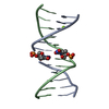
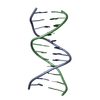
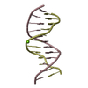
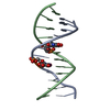
| Entry | Database: PDB / ID: 1d27 | ||||||||||||||||||
|---|---|---|---|---|---|---|---|---|---|---|---|---|---|---|---|---|---|---|---|
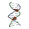
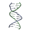
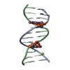
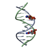
| Title | HIGH-RESOLUTION STRUCTURE OF A MUTAGENIC LESION IN DNA | ||||||||||||||||||
 Components Components | DNA (5'-D(* Keywords Keywords DNA / B-DNA / DOUBLE HELIX / MODIFIED / MISMATCHED DNA / B-DNA / DOUBLE HELIX / MODIFIED / MISMATCHEDFunction / homology | DNA / DNA (> 10) Function and homology information Function and homology informationMethod | X-RAY DIFFRACTION / Resolution: 2 Å  Authors AuthorsLeonard, G.A. / Thomson, J. / Watson, W.P. / Brown, T. |  CitationJournal: Proc.Natl.Acad.Sci.USA / Year: 1990 CitationJournal: Proc.Natl.Acad.Sci.USA / Year: 1990Title: High-resolution structure of a mutagenic lesion in DNA. Authors: Leonard, G.A. / Thomson, J. / Watson, W.P. / Brown, T. History |
|
- Structure visualization
| Structure viewer | Molecule: MolmilJmol/JSmol |
|---|
- Downloads & links
-Download
| PDBx/mmCIF format | 1d27.cif.gz | 25 KB | Display | PDBx/mmCIF format |
|---|---|---|---|---|
| PDB format | pdb1d27.ent.gz | 15.9 KB | Display | PDB format |
| PDBx/mmJSON format | 1d27.json.gz | Tree view | PDBx/mmJSON format | |
| Others |  Other downloads Other downloads |
-Validation report
| Arichive directory | https://data.pdbj.org/pub/pdb/validation_reports/d2/1d27ftp://data.pdbj.org/pub/pdb/validation_reports/d2/1d27 | HTTPS FTP |
|---|
-Related structure data
| Similar structure data |
|---|


-Links
 PDBj
PDBj


- Assembly
| Deposited unit | 
| ||||||||
|---|---|---|---|---|---|---|---|---|---|
| 1 |
| ||||||||
| Unit cell |
|
-Components
| #1: DNA chain | Mass: 3692.430 Da / Num. of mol.: 2 / Source method: obtained synthetically #2: Water | ChemComp-HOH / | Water Mass: 18.015 Da / Num. of mol.: 69 / Source method: isolated from a natural source / Formula: H2O Mass: 18.015 Da / Num. of mol.: 69 / Source method: isolated from a natural source / Formula: H2O |
|---|
-Experimental details
-Experiment
| Experiment | Method: X-RAY DIFFRACTION |
|---|
- Sample preparation
| Crystal | Density Matthews: 2.31 Å3/Da / Density % sol: 46.82 % | ||||||||||||||||||||||||||||||||||||
|---|---|---|---|---|---|---|---|---|---|---|---|---|---|---|---|---|---|---|---|---|---|---|---|---|---|---|---|---|---|---|---|---|---|---|---|---|---|
| Crystal grow | Temperature: 277 K / Method: vapor diffusion / pH: 6.3 / Details: pH 6.30, VAPOR DIFFUSION, temperature 277.00K | ||||||||||||||||||||||||||||||||||||
| Components of the solutions |
| ||||||||||||||||||||||||||||||||||||
| Crystal grow | *PLUS Temperature: 277 K / pH: 6.3 / Method: vapor diffusion | ||||||||||||||||||||||||||||||||||||
| Components of the solutions | *PLUS
|
-Data collection
| Diffraction |
| ||||||||||||
|---|---|---|---|---|---|---|---|---|---|---|---|---|---|
| Diffraction source |
| ||||||||||||
| Detector |
| ||||||||||||
| Radiation |
| ||||||||||||
| Radiation wavelength |
| ||||||||||||
| Reflection | Highest resolution: 2 Å / Num. obs: 4557 | ||||||||||||
| Reflection | *PLUS Highest resolution: 2 Å / % possible obs: 91 % / Rmerge(I) obs: 0.03 |
- Processing
| Software | Name: NUCLSQ / Classification: refinement | |||||||||||||||||||||||||||||||||||||||||||||||||||||||||||||||
|---|---|---|---|---|---|---|---|---|---|---|---|---|---|---|---|---|---|---|---|---|---|---|---|---|---|---|---|---|---|---|---|---|---|---|---|---|---|---|---|---|---|---|---|---|---|---|---|---|---|---|---|---|---|---|---|---|---|---|---|---|---|---|---|---|
| Refinement | Resolution: 2→7 Å / σ(F): 3 /
| |||||||||||||||||||||||||||||||||||||||||||||||||||||||||||||||
| Refine Biso |
| |||||||||||||||||||||||||||||||||||||||||||||||||||||||||||||||
| Refinement step | Cycle: LAST / Resolution: 2→7 Å
| |||||||||||||||||||||||||||||||||||||||||||||||||||||||||||||||
| Refine LS restraints |
| |||||||||||||||||||||||||||||||||||||||||||||||||||||||||||||||
| Refinement | *PLUS Highest resolution: 2 Å / Lowest resolution: 7 Å / σ(F): 3 / Rfactor obs: 0.185 / Num. reflection obs: 3118 | |||||||||||||||||||||||||||||||||||||||||||||||||||||||||||||||
| Solvent computation | *PLUS | |||||||||||||||||||||||||||||||||||||||||||||||||||||||||||||||
| Displacement parameters | *PLUS |
