 Movie
Movie Controller


+ Open data

- Basic information
| Entry | Database: PDB / ID: 1cjy | ||||||
|---|---|---|---|---|---|---|---|
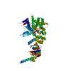
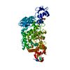
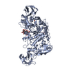
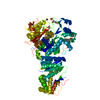
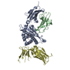
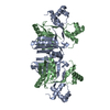
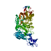
| Title | HUMAN CYTOSOLIC PHOSPHOLIPASE A2 | ||||||
 Components Components | PROTEIN (CYTOSOLIC PHOSPHOLIPASE A2) | ||||||
 Keywords Keywords |  HYDROLASE / PHOSPHOLIPASE / LIPID-BINDING HYDROLASE / PHOSPHOLIPASE / LIPID-BINDING | ||||||
| Function / homology |  Function and homology information Function and homology informationplatelet activating factor biosynthetic process / phosphatidylglycerol catabolic process / Arachidonic acid metabolism / icosanoid metabolic process / Acyl chain remodeling of CL / monoacylglycerol biosynthetic process / glycerophospholipid catabolic process / Hydrolysis of LPC / phosphatidyl phospholipase B activity / lysophospholipase ...platelet activating factor biosynthetic process / phosphatidylglycerol catabolic process / Arachidonic acid metabolism / icosanoid metabolic process / Acyl chain remodeling of CL / monoacylglycerol biosynthetic process / glycerophospholipid catabolic process / Hydrolysis of LPC / phosphatidyl phospholipase B activity / lysophospholipase / O-acyltransferase activity / phosphatidylcholine acyl-chain remodeling / Acyl chain remodelling of PG / calcium-independent phospholipase A2 activity / Acyl chain remodelling of PC / Acyl chain remodelling of PI / Acyl chain remodelling of PS / Acyl chain remodelling of PE / ceramide 1-phosphate binding / Synthesis of PA / glycerol metabolic process / arachidonic acid metabolic process / phosphatidylinositol-5-phosphate binding / positive regulation of T-helper 1 type immune response / phosphatidylcholine catabolic process / lysophospholipase activity / positive regulation of prostaglandin secretion / phospho-PLA2 pathway / calcium-dependent phospholipase A2 activity / COPI-independent Golgi-to-ER retrograde traffic / phosphatidylinositol-3-phosphate binding / positive regulation of macrophage activation / leukotriene biosynthetic process / calcium-dependent phospholipid binding / positive regulation of platelet activation / phosphatidylinositol-4-phosphate binding / phospholipase A2 activity / phospholipase A2 / prostaglandin biosynthetic process / cellular response to antibiotic / arachidonic acid secretion / Platelet sensitization by LDL / establishment of localization in cell / ADP signalling through P2Y purinoceptor 1 / nuclear envelope / regulation of cell population proliferation / mitochondrial inner membrane / Golgi membrane / intracellular membrane-bounded organelle / calcium ion binding / endoplasmic reticulum membrane / Golgi apparatus / endoplasmic reticulum / nucleus / cytosol / cytoplasmSimilarity search - Function | ||||||
| Biological species |  Homo sapiens (human) Homo sapiens (human) | ||||||
| Method | X-RAY DIFFRACTION / SYNCHROTRON / MAD / Resolution: 2.5 Å | ||||||
 Authors Authors | Dessen, A. / Tang, J. / Schmidt, H. / Stahl, M. / Clark, J.D. / Seehra, J. / Somers, W.S. | ||||||
 Citation Citation | Journal: Cell(Cambridge,Mass.) / Year: 1999 Title: Crystal structure of human cytosolic phospholipase A2 reveals a novel topology and catalytic mechanism. Authors: Dessen, A. / Tang, J. / Schmidt, H. / Stahl, M. / Clark, J.D. / Seehra, J. / Somers, W.S. | ||||||
| History |
|
- Structure visualization
| Structure viewer | Molecule: MolmilJmol/JSmol |
|---|
- Downloads & links
-Download
| PDBx/mmCIF format | 1cjy.cif.gz | 256.2 KB | Display | PDBx/mmCIF format |
|---|---|---|---|---|
| PDB format | pdb1cjy.ent.gz | 201.6 KB | Display | PDB format |
| PDBx/mmJSON format | 1cjy.json.gz | Tree view | PDBx/mmJSON format | |
| Others |  Other downloads Other downloads |
-Validation report
| Arichive directory | https://data.pdbj.org/pub/pdb/validation_reports/cj/1cjyftp://data.pdbj.org/pub/pdb/validation_reports/cj/1cjy | HTTPS FTP |
|---|
-Related structure data
| Similar structure data |
|---|


-Links
 PDBj
PDBj


- Assembly
| Deposited unit | 
| ||||||||
|---|---|---|---|---|---|---|---|---|---|
| 1 | 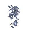
| ||||||||
| 2 | 
| ||||||||
| Unit cell |
|
-Components
| #1: Protein | Mass: 85309.734 Da / Num. of mol.: 2 Source method: isolated from a genetically manipulated source Source: (gene. exp.) Homo sapiens (human) / Production host:   Cricetulus griseus (Chinese hamster) / References: UniProt: P47712, phospholipase A2 Cricetulus griseus (Chinese hamster) / References: UniProt: P47712, phospholipase A2#2: Chemical | ChemComp-CA /   Mass: 40.078 Da / Num. of mol.: 4 / Source method: obtained synthetically / Formula: Ca Mass: 40.078 Da / Num. of mol.: 4 / Source method: obtained synthetically / Formula: Ca#3: Chemical | MES (buffer)  Mass: 195.237 Da / Num. of mol.: 2 / Source method: obtained synthetically / Formula: C6H13NO4S / Comment: pH buffer*YM Mass: 195.237 Da / Num. of mol.: 2 / Source method: obtained synthetically / Formula: C6H13NO4S / Comment: pH buffer*YM#4: Water | ChemComp-HOH / | Water Mass: 18.015 Da / Num. of mol.: 58 / Source method: isolated from a natural source / Formula: H2O Mass: 18.015 Da / Num. of mol.: 58 / Source method: isolated from a natural source / Formula: H2O |
|---|
-Experimental details
-Experiment
| Experiment | Method: X-RAY DIFFRACTION / Number of used crystals: 2 |
|---|
- Sample preparation
| Crystal | Density Matthews: 2.99 Å3/Da / Density % sol: 58.84 % | ||||||||||||||||||||||||||||||
|---|---|---|---|---|---|---|---|---|---|---|---|---|---|---|---|---|---|---|---|---|---|---|---|---|---|---|---|---|---|---|---|
| Crystal grow | pH: 8.5 / Details: pH 8.5 | ||||||||||||||||||||||||||||||
| Crystal | *PLUS Density % sol: 60 % | ||||||||||||||||||||||||||||||
| Crystal grow | *PLUS Temperature: 18 ℃ / pH: 6.2 / Method: vapor diffusion | ||||||||||||||||||||||||||||||
| Components of the solutions | *PLUS
|
-Data collection
| Diffraction | Mean temperature: 100 K | |||||||||
|---|---|---|---|---|---|---|---|---|---|---|
| Diffraction source | Source: SYNCHROTRON / Site: ALS  / Beamline: 5.0.2 / Wavelength: 1.2, 1.6496 / Beamline: 5.0.2 / Wavelength: 1.2, 1.6496 | |||||||||
| Detector | Type: ADSC / Detector: CCD / Date: Mar 1, 1998 | |||||||||
| Radiation | Protocol: MAD / Monochromatic (M) / Laue (L): M / Scattering type: x-ray | |||||||||
| Radiation wavelength |
| |||||||||
| Reflection | Resolution: 2.5→12 Å / Num. obs: 66223 / % possible obs: 93.3 % / Redundancy: 4.1 % / Rsym value: 0.064 / Net I/σ(I): 18.2 | |||||||||
| Reflection | *PLUS Num. measured all: 271686 / Rmerge(I) obs: 0.064 | |||||||||
| Reflection shell | *PLUS % possible obs: 87.9 % / Rmerge(I) obs: 0.3 / Mean I/σ(I) obs: 3.3 |
- Processing
| Software |
| ||||||||||||||||||||||||||||||||||||||||||||||||||||||||||||
|---|---|---|---|---|---|---|---|---|---|---|---|---|---|---|---|---|---|---|---|---|---|---|---|---|---|---|---|---|---|---|---|---|---|---|---|---|---|---|---|---|---|---|---|---|---|---|---|---|---|---|---|---|---|---|---|---|---|---|---|---|---|
| Refinement | Method to determine structure: MAD / Resolution: 2.5→12 Å / Cross valid method: THROUGHOUT / σ(F): 2
| ||||||||||||||||||||||||||||||||||||||||||||||||||||||||||||
| Refinement step | Cycle: LAST / Resolution: 2.5→12 Å
| ||||||||||||||||||||||||||||||||||||||||||||||||||||||||||||
| Refine LS restraints |
| ||||||||||||||||||||||||||||||||||||||||||||||||||||||||||||
| Software | *PLUS Name: X-PLOR / Version: 3.843 / Classification: refinement | ||||||||||||||||||||||||||||||||||||||||||||||||||||||||||||
| Refinement | *PLUS Highest resolution: 2.5 Å / Lowest resolution: 12 Å / σ(F): 2 / % reflection Rfree: 10 % | ||||||||||||||||||||||||||||||||||||||||||||||||||||||||||||
| Solvent computation | *PLUS | ||||||||||||||||||||||||||||||||||||||||||||||||||||||||||||
| Displacement parameters | *PLUS Biso mean: 47.7 Å2 |
