 Movie
Movie Controller

[English] 日本語
 Yorodumi
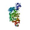
- PDB-2e8z: Crystal structure of pullulanase type I from Bacillus subtilis st... -
+ Open data

- Basic information
| Entry | Database: PDB / ID: 2e8z | |||||||||
|---|---|---|---|---|---|---|---|---|---|---|
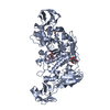
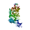
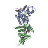
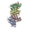
| Title | Crystal structure of pullulanase type I from Bacillus subtilis str. 168 complexed with alpha-cyclodextrin | |||||||||
 Components Components | AmyX protein | |||||||||
 Keywords Keywords | HYDROLASE / multiple domain / beta-alpha-barrel / alpha-amylase-family / alpha-cyclodextrin | |||||||||
| Function / homology |  Function and homology information Function and homology information | |||||||||
| Biological species |  Bacillus subtilis (bacteria) Bacillus subtilis (bacteria) | |||||||||
| Method | X-RAY DIFFRACTION / SYNCHROTRON / MOLECULAR REPLACEMENT / Resolution: 2.2 Å | |||||||||
 Authors Authors | Mikami, B. / Malle, D. / Utsumi, S. / Iwamoto, H. / Katsuya, Y. | |||||||||
 Citation Citation | Journal: To be Published Title: Crystal structure of pullulanase type I from Bacillus subtilis str. 168 in complex with maltose and alpha-cyclodextrin Authors: Malle, D. / Iwamoto, H. / Katsuya, Y. / Utsumi, S. / Mikami, B. #1: Journal: ACTA CRYSTALLOGR.,SECT.F / Year: 2006 Title: Overexpression, purification and preliminary X-ray analysis of pullulanase from Bacillus subtilis strain 168 Authors: Malle, D. / Itoh, T. / Hashimoto, W. / Murata, K. / Utsumi, S. / Mikami, B. | |||||||||
| History |
|
- Structure visualization
| Structure viewer | Molecule: MolmilJmol/JSmol |
|---|
- Downloads & links
-Download
| PDBx/mmCIF format | 2e8z.cif.gz | 306.3 KB | Display | PDBx/mmCIF format |
|---|---|---|---|---|
| PDB format | pdb2e8z.ent.gz | 246.4 KB | Display | PDB format |
| PDBx/mmJSON format | 2e8z.json.gz | Tree view | PDBx/mmJSON format | |
| Others |  Other downloads Other downloads |
-Validation report
| Arichive directory | https://data.pdbj.org/pub/pdb/validation_reports/e8/2e8zftp://data.pdbj.org/pub/pdb/validation_reports/e8/2e8z | HTTPS FTP |
|---|
-Related structure data
| Related structure data | 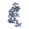 2e8yC  2e9bC 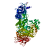 2fgzS C: citing same article ( S: Starting model for refinement |
|---|---|
| Similar structure data |


-Links
 PDBj
PDBj

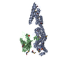
- Assembly
| Deposited unit | 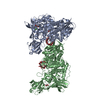
| ||||||||
|---|---|---|---|---|---|---|---|---|---|
| 1 | 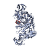
| ||||||||
| 2 | 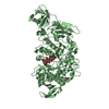
| ||||||||
| Unit cell |
|
-Components
-Protein / Sugars , 2 types, 4 molecules AB
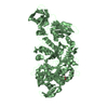
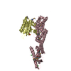
| #1: Protein | / Pullulanase Mass: 81206.703 Da / Num. of mol.: 2 Source method: isolated from a genetically manipulated source Source: (gene. exp.) Bacillus subtilis (bacteria) / Strain: 168 / Gene: amyX / Plasmid: pET21d / Production host: Escherichia coli (E. coli) / Strain (production host): HMS 174References: UniProt: O34587, UniProt: C0SPA0*PLUS, pullulanase#2: Polysaccharide | Α-Cyclodextrin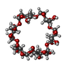  , Oligosaccharide / Class: Drug delivery / Mass: 990.860 Da / Num. of mol.: 2 , Oligosaccharide / Class: Drug delivery / Mass: 990.860 Da / Num. of mol.: 2Source method: isolated from a genetically manipulated source Details: cyclic oligosaccharide / References: alpha-cyclodextrin |
|---|
-Non-polymers , 4 types, 673 molecules 


| #3: Chemical |  Mass: 40.078 Da / Num. of mol.: 2 / Source method: obtained synthetically / Formula: Ca Mass: 40.078 Da / Num. of mol.: 2 / Source method: obtained synthetically / Formula: Ca#4: Chemical | Acetate Mass: 59.044 Da / Num. of mol.: 2 / Source method: obtained synthetically / Formula: C2H3O2 Mass: 59.044 Da / Num. of mol.: 2 / Source method: obtained synthetically / Formula: C2H3O2#5: Chemical | Glycerol Mass: 92.094 Da / Num. of mol.: 2 / Source method: obtained synthetically / Formula: C3H8O3 Mass: 92.094 Da / Num. of mol.: 2 / Source method: obtained synthetically / Formula: C3H8O3#6: Water | ChemComp-HOH / | WaterMass: 18.015 Da / Num. of mol.: 667 / Source method: isolated from a natural source / Formula: H2O |
|---|
-Experimental details
-Experiment
| Experiment | Method: X-RAY DIFFRACTION / Number of used crystals: 1 |
|---|
- Sample preparation
| Crystal | Density Matthews: 2.62 Å3/Da / Density % sol: 53 % |
|---|---|
| Crystal grow | Temperature: 293 K / Method: vapor diffusion / pH: 5.2 Details: 10% PEG 4000, 0.1M sodium acetate buffer, 0.2M magnesium acetate, 20mM alpha-cyclodextrin, pH 5.2, VAPOR DIFFUSION, temperature 293K |
-Data collection
| Diffraction | Mean temperature: 100 K |
|---|---|
| Diffraction source | Source: SYNCHROTRON / Site: SPring-8  / Beamline: BL38B1 / Wavelength: 1 Å / Beamline: BL38B1 / Wavelength: 1 Å |
| Detector | Type: RIGAKU JUPITER 210 / Detector: CCD / Date: Jul 16, 2005 / Details: mirrors |
| Radiation | Monochromator: Si 111 CHANNEL / Protocol: SINGLE WAVELENGTH / Monochromatic (M) / Laue (L): M / Scattering type: x-ray |
| Radiation wavelength | Wavelength: 1 Å / Relative weight: 1 |
| Reflection | Resolution: 2.2→40 Å / Num. all: 87567 / Num. obs: 87392 / % possible obs: 99.8 % / Observed criterion σ(I): 1 / Redundancy: 4.7 % / Biso Wilson estimate: 18.1 Å2 / Rmerge(I) obs: 0.092 / Net I/σ(I): 5.9 |
| Reflection shell | Resolution: 2.2→2.28 Å / Redundancy: 4.2 % / Rmerge(I) obs: 0.424 / Num. unique all: 8610 / % possible all: 99.3 |
- Processing
| Software |
| ||||||||||||||||||||||||||||||||||||
|---|---|---|---|---|---|---|---|---|---|---|---|---|---|---|---|---|---|---|---|---|---|---|---|---|---|---|---|---|---|---|---|---|---|---|---|---|---|
| Refinement | Method to determine structure: MOLECULAR REPLACEMENT Starting model: PDB ENTRY 2FGZ Resolution: 2.2→14.97 Å / Rfactor Rfree error: 0.003 / Data cutoff high absF: 3696398.23 / Data cutoff low absF: 0 / Isotropic thermal model: RESTRAINED / Cross valid method: THROUGHOUT / σ(F): 0 / Stereochemistry target values: Engh & Huber
| ||||||||||||||||||||||||||||||||||||
| Solvent computation | Solvent model: FLAT MODEL / Bsol: 44.5008 Å2 / ksol: 0.380901 e/Å3 | ||||||||||||||||||||||||||||||||||||
| Displacement parameters | Biso mean: 27 Å2
| ||||||||||||||||||||||||||||||||||||
| Refine analyze |
| ||||||||||||||||||||||||||||||||||||
| Refinement step | Cycle: LAST / Resolution: 2.2→14.97 Å
| ||||||||||||||||||||||||||||||||||||
| Refine LS restraints |
| ||||||||||||||||||||||||||||||||||||
| LS refinement shell | Resolution: 2.2→2.34 Å / Rfactor Rfree error: 0.007 / Total num. of bins used: 6
| ||||||||||||||||||||||||||||||||||||
| Xplor file |
|
