 Movie
Movie Controller

[English] 日本語
 Yorodumi
- PDB-1chp: SURPRISING LEADS FOR A CHOLERA TOXIN RECEPTOR BINDING ANTAGONIST;... -
+ Open data

- Basic information
| Entry | Database: PDB / ID: 1chp | ||||||
|---|---|---|---|---|---|---|---|
| Title | SURPRISING LEADS FOR A CHOLERA TOXIN RECEPTOR BINDING ANTAGONIST; CRYSTALLOGRAPHIC STUDIES OF CTB MUTANTS | ||||||
 Components Components | CHOLERA TOXIN B PENTAMER | ||||||
 Keywords Keywords |  TOXIN TOXIN | ||||||
| Function / homology |  Function and homology information Function and homology informationhost cell surface binding / galactose binding / catalytic complex / positive regulation of tyrosine phosphorylation of STAT protein / toxin activity / periplasmic space / host cell plasma membrane / extracellular region / membraneSimilarity search - Function | ||||||
| Biological species |   Vibrio cholerae (bacteria) Vibrio cholerae (bacteria) | ||||||
| Method | X-RAY DIFFRACTION / Resolution: 2 Å | ||||||
 Authors Authors | Merritt, E.A. / Hol, W.G.J. | ||||||
 Citation Citation | Journal: Structure / Year: 1995 Title: Surprising leads for a cholera toxin receptor-binding antagonist: crystallographic studies of CTB mutants. Authors: Merritt, E.A. / Sarfaty, S. / Chang, T.T. / Palmer, L.M. / Jobling, M.G. / Holmes, R.K. / Hol, W.G. #1: Journal: Protein Sci. / Year: 1994Title: 2.2 Angstroms Crystal Structure of Cholera Toxin B5 Pentamer Bound to Receptor GM1 Pentasaccharide Authors: Merritt, E.A. / Sarfaty, S. / Van Den Akker, F. / L'Hoir, C. / Martial, J.A. / Hol, W.G.J. #2: Journal: Mol.Microbiol. / Year: 1991Title: Analysis of Structure and Function of the B Subunit of Cholera Toxin by the Use of Site-Directed Mutagenesis Authors: Jobling, M.G. / Holmes, R.K. | ||||||
| History |
| ||||||
| Remark 700 | SHEET IN THE PENTAMER THE BETA SHEETS FROM ADJACENT MONOMERS COMBINE TO FORM A CONTINUOUS SIX- ...SHEET IN THE PENTAMER THE BETA SHEETS FROM ADJACENT MONOMERS COMBINE TO FORM A CONTINUOUS SIX-STRANDED ANTI-PARALLEL SHEET ACROSS EACH MONOMER-MONOMER INTERFACE. |
- Structure visualization
| Structure viewer | Molecule: MolmilJmol/JSmol |
|---|
- Downloads & links
-Download
| PDBx/mmCIF format | 1chp.cif.gz | 110.8 KB | Display | PDBx/mmCIF format |
|---|---|---|---|---|
| PDB format | pdb1chp.ent.gz | 92.2 KB | Display | PDB format |
| PDBx/mmJSON format | 1chp.json.gz | Tree view | PDBx/mmJSON format | |
| Others |  Other downloads Other downloads |
-Validation report
| Arichive directory | https://data.pdbj.org/pub/pdb/validation_reports/ch/1chpftp://data.pdbj.org/pub/pdb/validation_reports/ch/1chp | HTTPS FTP |
|---|
-Related structure data


-Links
 PDBj
PDBj
- Assembly
| Deposited unit | 
| ||||||||
|---|---|---|---|---|---|---|---|---|---|
| 1 |
| ||||||||
| Unit cell |
| ||||||||
| Atom site foot note | 1: CIS PROLINE - PRO D 93 / 2: CIS PROLINE - PRO E 93 / 3: CIS PROLINE - PRO F 93 / 4: CIS PROLINE - PRO G 93 / 5: CIS PROLINE - PRO H 93 | ||||||||
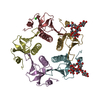
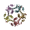
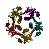
| Details | CHOLERA TOXIN IS AN AB5 HEXAMER. THE B-PENTAMER OF CHOLERA TOXIN IS RESPONSIBLE FOR RECEPTOR RECOGNITION AND BINDING. |
-Components
| #1: Protein | Mass: 11681.303 Da / Num. of mol.: 5 / Mutation: G33D Source method: isolated from a genetically manipulated source Source: (gene. exp.) Vibrio cholerae (bacteria) / Strain: OGAWA 41 (CLASSICAL) / Production host: Escherichia coli (E. coli) / References: UniProt: P01556#2: Chemical | ChemComp-CL / | Chloride  Mass: 35.453 Da / Num. of mol.: 1 / Source method: obtained synthetically / Formula: Cl Mass: 35.453 Da / Num. of mol.: 1 / Source method: obtained synthetically / Formula: Cl#3: Water | ChemComp-HOH / | Water Mass: 18.015 Da / Num. of mol.: 248 / Source method: isolated from a natural source / Formula: H2O Mass: 18.015 Da / Num. of mol.: 248 / Source method: isolated from a natural source / Formula: H2OCompound details | THE FIVE IDENTICAL B SUBUNITS ARE LABELLED AS CHAINS D, E, F, G, AND H CORRESPONDING TO CHAIN ...THE FIVE IDENTICAL B SUBUNITS ARE LABELLED AS CHAINS D, E, F, G, AND H CORRESPOND | |
|---|
-Experimental details
-Experiment
| Experiment | Method: X-RAY DIFFRACTION |
|---|
- Sample preparation
| Crystal | Density Matthews: 2.18 Å3/Da / Density % sol: 43.62 % | ||||||||||||||||||||||||||||||||||||
|---|---|---|---|---|---|---|---|---|---|---|---|---|---|---|---|---|---|---|---|---|---|---|---|---|---|---|---|---|---|---|---|---|---|---|---|---|---|
| Crystal grow | pH: 7.5 / Details: pH 7.5 | ||||||||||||||||||||||||||||||||||||
| Crystal grow | *PLUS Method: vapor diffusion, sitting drop | ||||||||||||||||||||||||||||||||||||
| Components of the solutions | *PLUS
|
-Data collection
| Diffraction source | Wavelength: 1.54 |
|---|---|
| Detector | Type: SIEMENS X1000 / Detector: AREA DETECTOR / Date: Jul 14, 1994 |
| Radiation | Monochromatic (M) / Laue (L): M / Scattering type: x-ray |
| Radiation wavelength | Wavelength: 1.54 Å / Relative weight: 1 |
| Reflection | Num. obs: 32710 / % possible obs: 99 % / Observed criterion σ(I): 0 / Redundancy: 2.85 % / Rmerge(I) obs: 0.081 |
| Reflection | *PLUS Highest resolution: 2 Å / Rmerge(I) obs: 0.081 |
- Processing
| Software |
| ||||||||||||||||||||||||||||||||||||||||||||||||||||||||||||
|---|---|---|---|---|---|---|---|---|---|---|---|---|---|---|---|---|---|---|---|---|---|---|---|---|---|---|---|---|---|---|---|---|---|---|---|---|---|---|---|---|---|---|---|---|---|---|---|---|---|---|---|---|---|---|---|---|---|---|---|---|---|
| Refinement | Resolution: 2→12 Å / σ(F): 3
| ||||||||||||||||||||||||||||||||||||||||||||||||||||||||||||
| Displacement parameters | Biso mean: 21.5 Å2 | ||||||||||||||||||||||||||||||||||||||||||||||||||||||||||||
| Refinement step | Cycle: LAST / Resolution: 2→12 Å
| ||||||||||||||||||||||||||||||||||||||||||||||||||||||||||||
| Refine LS restraints |
| ||||||||||||||||||||||||||||||||||||||||||||||||||||||||||||
| Software | *PLUS Name: X-PLOR / Classification: refinement | ||||||||||||||||||||||||||||||||||||||||||||||||||||||||||||
| Refinement | *PLUS | ||||||||||||||||||||||||||||||||||||||||||||||||||||||||||||
| Solvent computation | *PLUS | ||||||||||||||||||||||||||||||||||||||||||||||||||||||||||||
| Displacement parameters | *PLUS |
