 Movie
Movie Controller


+ Open data

- Basic information
| Entry | Database: PDB / ID: 1byl | ||||||
|---|---|---|---|---|---|---|---|
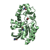
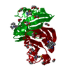
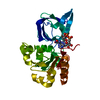
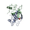
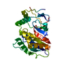
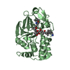
| Title | BLEOMYCIN RESISTANCE PROTEIN FROM STREPTOALLOTEICHUS HINDUSTANUS | ||||||
 Components Components | PROTEIN (BLEOMYCIN RESISTANCE PROTEIN) | ||||||
 Keywords Keywords |  ANTIBIOTIC / ANTIBIOTIC RESISTANCE / BLEOMYCIN / DRUG SEQUESTERING / CHAIN SWAPPING ANTIBIOTIC / ANTIBIOTIC RESISTANCE / BLEOMYCIN / DRUG SEQUESTERING / CHAIN SWAPPING | ||||||
| Function / homology |  Function and homology information Function and homology information | ||||||
| Biological species |  Streptoalloteichus hindustanus (bacteria) Streptoalloteichus hindustanus (bacteria) | ||||||
| Method | X-RAY DIFFRACTION / MIRAS / Resolution: 2.3 Å | ||||||
 Authors Authors | Dumas, P. / Bergdoll, M. / Cagnon, C. / Masson, J.M. | ||||||
 Citation Citation | Journal: EMBO J. / Year: 1994 Title: Crystal structure and site-directed mutagenesis of a bleomycin resistance protein and their significance for drug sequestering. Authors: Dumas, P. / Bergdoll, M. / Cagnon, C. / Masson, J.M. #1: Journal: Embo J. / Year: 1989Title: Crystallization and Preliminary X-Ray Data of a Phleomycin-Binding Protein from Streptoalloteichus Hindistanus Authors: Rondeau, J.M. / Cagnon, C. / Moras, D. / Masson, J.M. #2: Journal: FEBS Lett. / Year: 1988Title: Bleomycin Resistance Conferred by a Drug-Binding Protein Authors: Gatignol, A. / Durand, H. / Tiraby, G. #3: Journal: Protein Sci. / Year: 1998Title: All in the Family: Structural and Evolutionary Relationships Among Three Modular Proteins with Functions and Variable Assembly Authors: Bergdoll, M. / Eltis, L.D. / Cameron, A.D. / Dumas, P. / Bolin, J.T. | ||||||
| History |
|
- Structure visualization
| Structure viewer | Molecule: MolmilJmol/JSmol |
|---|
- Downloads & links
-Download
| PDBx/mmCIF format | 1byl.cif.gz | 37.8 KB | Display | PDBx/mmCIF format |
|---|---|---|---|---|
| PDB format | pdb1byl.ent.gz | 26.1 KB | Display | PDB format |
| PDBx/mmJSON format | 1byl.json.gz | Tree view | PDBx/mmJSON format | |
| Others |  Other downloads Other downloads |
-Validation report
| Arichive directory | https://data.pdbj.org/pub/pdb/validation_reports/by/1bylftp://data.pdbj.org/pub/pdb/validation_reports/by/1byl | HTTPS FTP |
|---|
-Related structure data
| Similar structure data |
|---|


-Links
 PDBj
PDBj- Assembly
| Deposited unit | 
| ||||||||
|---|---|---|---|---|---|---|---|---|---|
| 1 | 
| ||||||||
| Unit cell |
| ||||||||
| Details | THE PROTEIN'S COORDINATES IN THIS FILE CORRESPOND TO HALF OF THE FUNCTIONNAL UNIT THE ACTIVE MOLECULE IS MADE UP OF TWO TIGHTLY INTERACTING MONOMERS WHICH ARE INVOLVED IN MUTUAL ARM EXCHANGE. |
-Components
| #1: Protein | Mass: 13953.388 Da / Num. of mol.: 1 Source method: isolated from a genetically manipulated source Source: (gene. exp.) Streptoalloteichus hindustanus (bacteria)Description: DROCOURT, D., CALMELS, T., REYNES, J.P., BARON, M. & TIRABY, G. (1990) NUCLEIC ACIDS RES., 18, 4009. Cellular location: CYTOPLASMIC Cytoplasm / Gene: SH BLE / Plasmid: PIN-III-OMPA2 / Cellular location (production host): PERIPLASM / Gene (production host): SH BLE / Production host: Escherichia coli (E. coli) / Strain (production host): SG936 / References: UniProt: P17493 |
|---|---|
| #2: Water | ChemComp-HOH / Water Mass: 18.015 Da / Num. of mol.: 62 / Source method: isolated from a natural source / Formula: H2O Mass: 18.015 Da / Num. of mol.: 62 / Source method: isolated from a natural source / Formula: H2O |
| Sequence details | THREE EXTRA RESIDUES (ALA -2, GLU -1 & PHE 0) ARE PRESENT AT THE N-TERMINUS AND ARE PART OF AN ...THREE EXTRA RESIDUES (ALA -2, GLU -1 & PHE 0) ARE PRESENT AT THE N-TERMINUS AND ARE PART OF AN INCOMPLETE |
-Experimental details
-Experiment
| Experiment | Method: X-RAY DIFFRACTION / Number of used crystals: 1 |
|---|
- Sample preparation
| Crystal | Density Matthews: 2.3 Å3/Da / Density % sol: 50 % Description: LOCHVAT IS A SCALING AND HEAVY ATOM SITE DETERMINATION PROGRAM BY P. DUMAS [ ACTA CRYST A50 (1994) 526-546] |
|---|---|
| Crystal grow | pH: 6 / Details: SEE REFERENCE 2, pH 6.0 |
| Crystal grow | *PLUS Method: other / Details: Rondeau, J.M., (1989) J. Mol. Biol., 207, 645. |
-Data collection
| Diffraction | Mean temperature: 293 K |
|---|---|
| Diffraction source | Source: ROTATING ANODE / Type: SIEMENS / Wavelength: 1.5418 |
| Detector | Type: SIEMENS / Detector: AREA DETECTOR / Date: Apr 15, 1991 |
| Radiation | Monochromator: NI FILTER / Protocol: SINGLE WAVELENGTH / Monochromatic (M) / Laue (L): M / Scattering type: x-ray |
| Radiation wavelength | Wavelength: 1.5418 Å / Relative weight: 1 |
| Reflection | Resolution: 2.3→44.5 Å / Num. obs: 5959 / % possible obs: 93.3 % / Observed criterion σ(I): 2 / Redundancy: 8.3 % / Rsym value: 0.669 / Net I/σ(I): 48.4 |
| Reflection shell | Resolution: 2.3→2.44 Å / Redundancy: 3.4 % / Mean I/σ(I) obs: 4.24 / Rsym value: 0.204 / % possible all: 58 |
| Reflection | *PLUS Num. measured all: 49546 / Rmerge(I) obs: 0.067 |
- Processing
| Software |
| ||||||||||||||||||||||||||||||||||||||||||||||||||||||||||||
|---|---|---|---|---|---|---|---|---|---|---|---|---|---|---|---|---|---|---|---|---|---|---|---|---|---|---|---|---|---|---|---|---|---|---|---|---|---|---|---|---|---|---|---|---|---|---|---|---|---|---|---|---|---|---|---|---|---|---|---|---|---|
| Refinement | Method to determine structure: MIRAS / Resolution: 2.3→8 Å / Data cutoff high absF: 1000000 / Data cutoff low absF: 0.001 Cross valid method: ANOMALOUS DIFFERENCE OF THE NATIVE DATA SET ALLOWING TO LOCATE ALL SULFUR ATOMS σ(F): 2
| ||||||||||||||||||||||||||||||||||||||||||||||||||||||||||||
| Displacement parameters | Biso mean: 17.9 Å2 | ||||||||||||||||||||||||||||||||||||||||||||||||||||||||||||
| Refine analyze | Luzzati coordinate error obs: 0.25 Å / Luzzati d res low obs: 8 Å | ||||||||||||||||||||||||||||||||||||||||||||||||||||||||||||
| Refinement step | Cycle: LAST / Resolution: 2.3→8 Å
| ||||||||||||||||||||||||||||||||||||||||||||||||||||||||||||
| Refine LS restraints |
| ||||||||||||||||||||||||||||||||||||||||||||||||||||||||||||
| LS refinement shell | Resolution: 2.3→2.38 Å / Total num. of bins used: 10
| ||||||||||||||||||||||||||||||||||||||||||||||||||||||||||||
| Xplor file |
| ||||||||||||||||||||||||||||||||||||||||||||||||||||||||||||
| Software | *PLUS Name: X-PLOR / Version: 2.1 / Classification: refinement | ||||||||||||||||||||||||||||||||||||||||||||||||||||||||||||
| Refinement | *PLUS Highest resolution: 2.3 Å / Lowest resolution: 8 Å / σ(F): 2 | ||||||||||||||||||||||||||||||||||||||||||||||||||||||||||||
| Solvent computation | *PLUS | ||||||||||||||||||||||||||||||||||||||||||||||||||||||||||||
| Displacement parameters | *PLUS Biso mean: 17.9 Å2 | ||||||||||||||||||||||||||||||||||||||||||||||||||||||||||||
| Refine LS restraints | *PLUS Type: x_angle_deg / Dev ideal: 3.2 | ||||||||||||||||||||||||||||||||||||||||||||||||||||||||||||
| LS refinement shell | *PLUS % reflection Rfree: 21 % / Rfactor Rwork: 0.283 |
