 Movie
Movie Controller

[English] 日本語
 Yorodumi
- PDB-4iag: Crystal structure of ZbmA, the zorbamycin binding protein from St... -
+ Open data

- Basic information
| Entry | Database: PDB / ID: 4iag | ||||||
|---|---|---|---|---|---|---|---|
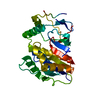
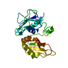
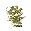
| Title | Crystal structure of ZbmA, the zorbamycin binding protein from Streptomyces flavoviridis | ||||||
 Components Components | Zbm binding protein | ||||||
 Keywords Keywords | Zorbamycin Binding Protein /  Structural Genomics / PSI-Biology / Midwest Center for Structural Genomics / MCSG / Enzyme Discovery for Natural Product Biosynthesis / NatPro / BLMA_like / zorbamycin / reductive isopropylation Structural Genomics / PSI-Biology / Midwest Center for Structural Genomics / MCSG / Enzyme Discovery for Natural Product Biosynthesis / NatPro / BLMA_like / zorbamycin / reductive isopropylation | ||||||
| Function / homology | Bleomycin resistance protein / 2,3-Dihydroxybiphenyl 1,2-Dioxygenase, domain 1 / 2,3-Dihydroxybiphenyl 1,2-Dioxygenase; domain 1 / Glyoxalase/Bleomycin resistance protein/Dihydroxybiphenyl dioxygenase / response to antibiotic / Roll / Alpha Beta / Zbm binding protein Function and homology information Function and homology information | ||||||
| Biological species |  Streptomyces flavoviridis (bacteria) Streptomyces flavoviridis (bacteria) | ||||||
| Method | X-RAY DIFFRACTION / SYNCHROTRON / SAD / Resolution: 1.9 Å | ||||||
 Authors Authors | Cuff, M.E. / Bigelow, L. / Bruno, C.J.P. / Clancy, S. / Babnigg, G. / Bingman, C.A. / Yennamalli, R. / Lohman, J. / Ma, M. / Shen, B. ...Cuff, M.E. / Bigelow, L. / Bruno, C.J.P. / Clancy, S. / Babnigg, G. / Bingman, C.A. / Yennamalli, R. / Lohman, J. / Ma, M. / Shen, B. / Phillips Jr., G.N. / Joachimiak, A. / Midwest Center for Structural Genomics (MCSG) / Enzyme Discovery for Natural Product Biosynthesis (NatPro) | ||||||
 Citation Citation | Journal: Biochemistry / Year: 2015 Title: Crystal Structure of the Zorbamycin-Binding Protein ZbmA, the Primary Self-Resistance Element in Streptomyces flavoviridis ATCC21892. Authors: Rudolf, J.D. / Bigelow, L. / Chang, C. / Cuff, M.E. / Lohman, J.R. / Chang, C.Y. / Ma, M. / Yang, D. / Clancy, S. / Babnigg, G. / Joachimiak, A. / Phillips, G.N. / Shen, B. | ||||||
| History |
|
- Structure visualization
| Structure viewer | Molecule: MolmilJmol/JSmol |
|---|
- Downloads & links
-Download
| PDBx/mmCIF format | 4iag.cif.gz | 63.8 KB | Display | PDBx/mmCIF format |
|---|---|---|---|---|
| PDB format | pdb4iag.ent.gz | 49.7 KB | Display | PDB format |
| PDBx/mmJSON format | 4iag.json.gz | Tree view | PDBx/mmJSON format | |
| Others |  Other downloads Other downloads |
-Validation report
| Arichive directory | https://data.pdbj.org/pub/pdb/validation_reports/ia/4iagftp://data.pdbj.org/pub/pdb/validation_reports/ia/4iag | HTTPS FTP |
|---|
-Related structure data


-Links
 PDBj
PDBj- Assembly
| Deposited unit | 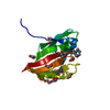
| ||||||||
|---|---|---|---|---|---|---|---|---|---|
| 1 | 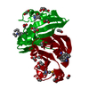
| ||||||||
| Unit cell |
| ||||||||
| Details | Likely forms a dimer with the crystallographic symmetry mate: x,y,z and 1-y, 1-x, 1/6-z. |
-Components
| #1: Protein | Mass: 15015.371 Da / Num. of mol.: 1 Source method: isolated from a genetically manipulated source Source: (gene. exp.) Streptomyces flavoviridis (bacteria) / Strain: ATCC 21892 / Gene: zbmA / Plasmid: pMCSG57 / Production host: Escherichia coli (E. coli) / Strain (production host): BL21(DE3)magic / References: UniProt: B9UIZ4 | ||||||
|---|---|---|---|---|---|---|---|
| #2: Chemical | ChemComp-EDO / Ethylene glycol  Mass: 62.068 Da / Num. of mol.: 8 / Source method: obtained synthetically / Formula: C2H6O2 Mass: 62.068 Da / Num. of mol.: 8 / Source method: obtained synthetically / Formula: C2H6O2#3: Chemical | ChemComp-GOL / | Glycerol  Mass: 92.094 Da / Num. of mol.: 1 / Source method: obtained synthetically / Formula: C3H8O3 Mass: 92.094 Da / Num. of mol.: 1 / Source method: obtained synthetically / Formula: C3H8O3#4: Water | ChemComp-HOH / | Water Mass: 18.015 Da / Num. of mol.: 55 / Source method: isolated from a natural source / Formula: H2O Mass: 18.015 Da / Num. of mol.: 55 / Source method: isolated from a natural source / Formula: H2OCompound details | THE LYSINE RESIDUES IN THIS PROTEIN WERE SUBJECT TO REDUCTIVE ISOPROPYLA | |
-Experimental details
-Experiment
| Experiment | Method: X-RAY DIFFRACTION / Number of used crystals: 1 |
|---|
- Sample preparation
| Crystal | Density Matthews: 1.97 Å3/Da / Density % sol: 37.45 % |
|---|---|
| Crystal grow | Temperature: 289 K / Method: vapor diffusion, sitting drop / pH: 4 Details: protein subject to reductive isopropylation, 0.8M Ammonium Sulfate, 0.1M Sodium Sitrate: HCl pH 4, VAPOR DIFFUSION, SITTING DROP, temperature 289K |
-Data collection
| Diffraction | Mean temperature: 100 K | |||||||||||||||||||||||||||||||||||||||||||||||||||||||||||||||||||||||||||||||||||||||||||||||||||||||||||||||||||||||||||||||||||||||||||||||||||
|---|---|---|---|---|---|---|---|---|---|---|---|---|---|---|---|---|---|---|---|---|---|---|---|---|---|---|---|---|---|---|---|---|---|---|---|---|---|---|---|---|---|---|---|---|---|---|---|---|---|---|---|---|---|---|---|---|---|---|---|---|---|---|---|---|---|---|---|---|---|---|---|---|---|---|---|---|---|---|---|---|---|---|---|---|---|---|---|---|---|---|---|---|---|---|---|---|---|---|---|---|---|---|---|---|---|---|---|---|---|---|---|---|---|---|---|---|---|---|---|---|---|---|---|---|---|---|---|---|---|---|---|---|---|---|---|---|---|---|---|---|---|---|---|---|---|---|---|---|
| Diffraction source | Source: SYNCHROTRON / Site: APS  / Beamline: 19-BM / Wavelength: 0.97929 Å / Beamline: 19-BM / Wavelength: 0.97929 Å | |||||||||||||||||||||||||||||||||||||||||||||||||||||||||||||||||||||||||||||||||||||||||||||||||||||||||||||||||||||||||||||||||||||||||||||||||||
| Detector | Type: ADSC QUANTUM 210r / Detector: CCD / Date: Aug 3, 2012 | |||||||||||||||||||||||||||||||||||||||||||||||||||||||||||||||||||||||||||||||||||||||||||||||||||||||||||||||||||||||||||||||||||||||||||||||||||
| Radiation | Monochromator: SAGITALLY FOCUSED Si(111) / Protocol: SAD / Monochromatic (M) / Laue (L): M / Scattering type: x-ray | |||||||||||||||||||||||||||||||||||||||||||||||||||||||||||||||||||||||||||||||||||||||||||||||||||||||||||||||||||||||||||||||||||||||||||||||||||
| Radiation wavelength | Wavelength: 0.97929 Å / Relative weight: 1 | |||||||||||||||||||||||||||||||||||||||||||||||||||||||||||||||||||||||||||||||||||||||||||||||||||||||||||||||||||||||||||||||||||||||||||||||||||
| Reflection | Redundancy: 9.7 % / Av σ(I) over netI: 33.01 / Number: 101644 / Rmerge(I) obs: 0.068 / Χ2: 1.23 / D res high: 1.9 Å / D res low: 50 Å / Num. obs: 10484 / % possible obs: 98.5 | |||||||||||||||||||||||||||||||||||||||||||||||||||||||||||||||||||||||||||||||||||||||||||||||||||||||||||||||||||||||||||||||||||||||||||||||||||
| Diffraction reflection shell |
| |||||||||||||||||||||||||||||||||||||||||||||||||||||||||||||||||||||||||||||||||||||||||||||||||||||||||||||||||||||||||||||||||||||||||||||||||||
| Reflection | Resolution: 1.9→50 Å / Num. all: 10484 / Num. obs: 10484 / % possible obs: 98.5 % / Observed criterion σ(I): -3 / Redundancy: 9.7 % / Biso Wilson estimate: 28.2 Å2 / Rmerge(I) obs: 0.068 / Χ2: 1.225 / Net I/σ(I): 11.2 | |||||||||||||||||||||||||||||||||||||||||||||||||||||||||||||||||||||||||||||||||||||||||||||||||||||||||||||||||||||||||||||||||||||||||||||||||||
| Reflection shell |
|
-Phasing
| Phasing | Method: SAD | ||||||||||||||||||||||||||||||||||||||||||||||||||||||||||||||||||||||||||||||||||||
|---|---|---|---|---|---|---|---|---|---|---|---|---|---|---|---|---|---|---|---|---|---|---|---|---|---|---|---|---|---|---|---|---|---|---|---|---|---|---|---|---|---|---|---|---|---|---|---|---|---|---|---|---|---|---|---|---|---|---|---|---|---|---|---|---|---|---|---|---|---|---|---|---|---|---|---|---|---|---|---|---|---|---|---|---|---|
| Phasing MAD | D res high: 2 Å / D res low: 44.75 Å / FOM : 0.172 / FOM acentric: 0.248 / FOM centric: 0 / Reflection: 9000 / Reflection acentric: 6258 / Reflection centric: 2742 | ||||||||||||||||||||||||||||||||||||||||||||||||||||||||||||||||||||||||||||||||||||
| Phasing MAD set | R cullis acentric: 2.07 / R cullis centric: 1 / Highest resolution: 2 Å / Lowest resolution: 44.75 Å / Loc acentric: 0 / Loc centric: 0 / Power acentric: 0 / Power centric: 0 / Reflection acentric: 6258 / Reflection centric: 2742 | ||||||||||||||||||||||||||||||||||||||||||||||||||||||||||||||||||||||||||||||||||||
| Phasing MAD set shell | ID: 1 / R cullis centric: 1 / Power acentric: 0 / Power centric: 0
| ||||||||||||||||||||||||||||||||||||||||||||||||||||||||||||||||||||||||||||||||||||
| Phasing MAD set site | Atom type symbol: Se / B iso: 53.9197 / Fract x: 0.914 / Fract y: 0.714 / Fract z: 0.017 / Occupancy: 4.23 / Occupancy iso: 0 | ||||||||||||||||||||||||||||||||||||||||||||||||||||||||||||||||||||||||||||||||||||
| Phasing MAD shell |
| ||||||||||||||||||||||||||||||||||||||||||||||||||||||||||||||||||||||||||||||||||||
| Phasing dm | Method: Solvent flattening and Histogram matching / Reflection: 10313 | ||||||||||||||||||||||||||||||||||||||||||||||||||||||||||||||||||||||||||||||||||||
| Phasing dm shell |
|
- Processing
| Software |
| ||||||||||||||||||||||||||||||||||||||||||||||||||||||||||||||||||||||||
|---|---|---|---|---|---|---|---|---|---|---|---|---|---|---|---|---|---|---|---|---|---|---|---|---|---|---|---|---|---|---|---|---|---|---|---|---|---|---|---|---|---|---|---|---|---|---|---|---|---|---|---|---|---|---|---|---|---|---|---|---|---|---|---|---|---|---|---|---|---|---|---|---|---|
| Refinement | Method to determine structure: SAD / Resolution: 1.9→32.59 Å / Cor.coef. Fo:Fc: 0.959 / Cor.coef. Fo:Fc free: 0.948 / WRfactor Rfree: 0.2317 / WRfactor Rwork: 0.1832 / Occupancy max: 1 / Occupancy min: 0.5 / FOM work R set: 0.8678 / SU B: 7.7 / SU ML: 0.107 / SU R Cruickshank DPI: 0.1613 / SU Rfree: 0.1483 / Cross valid method: THROUGHOUT / σ(F): 0 / ESU R: 0.161 / ESU R Free: 0.148 Stereochemistry target values: MAXIMUM LIKELIHOOD WITH PHASES Details: HYDROGENS HAVE BEEN ADDED IN THE RIDING POSITIONS U VALUES : WITH TLS ADDED
| ||||||||||||||||||||||||||||||||||||||||||||||||||||||||||||||||||||||||
| Solvent computation | Ion probe radii: 0.8 Å / Shrinkage radii: 0.8 Å / VDW probe radii: 1.2 Å / Solvent model: MASK | ||||||||||||||||||||||||||||||||||||||||||||||||||||||||||||||||||||||||
| Displacement parameters | Biso max: 97.97 Å2 / Biso mean: 38.2635 Å2 / Biso min: 20.07 Å2
| ||||||||||||||||||||||||||||||||||||||||||||||||||||||||||||||||||||||||
| Refinement step | Cycle: LAST / Resolution: 1.9→32.59 Å
| ||||||||||||||||||||||||||||||||||||||||||||||||||||||||||||||||||||||||
| Refine LS restraints |
| ||||||||||||||||||||||||||||||||||||||||||||||||||||||||||||||||||||||||
| LS refinement shell | Resolution: 1.9→1.949 Å / Total num. of bins used: 20
| ||||||||||||||||||||||||||||||||||||||||||||||||||||||||||||||||||||||||
| Refinement TLS params. | Method: refined / Origin x: 46.1737 Å / Origin y: -2.9324 Å / Origin z: 32.3225 Å
|
