non-canonical NF-kappaB signal transduction / canonical NF-kappaB signal transduction / response to cytokine / RNA polymerase II transcription regulatory region sequence-specific DNA binding / DNA-binding transcription factor activity, RNA polymerase II-specific / innate immune response / chromatin binding / negative regulation of transcription by RNA polymerase II / positive regulation of transcription by RNA polymerase II / nucleoplasm / cytosol Similarity search - Function
Type: MAR scanner 345 mm plate / Detector: IMAGE PLATE / Date: Oct 16, 1997 / Details: MIRRORS
Radiation
Monochromatic (M) / Laue (L): M / Scattering type: x-ray
Radiation wavelength
Wavelength: 0.997 Å / Relative weight: 1
Reflection
Resolution: 2.7→20 Å / Num. obs: 8927 / % possible obs: 82.9 % / Observed criterion σ(I): 0 / Redundancy: 3.8 % / Biso Wilson estimate: 93.5 Å2 / Rsym value: 0.041 / Net I/σ(I): 18.9
Reflection shell
Resolution: 2.7→2.8 Å / Redundancy: 2.8 % / Mean I/σ(I) obs: 2.7 / Rsym value: 0.286 / % possible all: 88.5
Reflection
*PLUS
Num. measured all: 33776 / Rmerge(I) obs: 0.041
Reflection shell
*PLUS
% possible obs: 88.5 % / Rmerge(I) obs: 0.286
-
Processing
Software
Name
Version
Classification
DENZO
datareduction
SCALEPACK
datascaling
X-PLOR
3.8
modelbuilding
X-PLOR
3.8
refinement
X-PLOR
3.8
phasing
Refinement
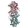
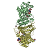
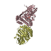
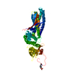
Method to determine structure: MIR / Resolution: 2.7→10 Å / Rfactor Rfree error: 0.014 / Isotropic thermal model: RESTRAINED / Cross valid method: THROUGHOUT / σ(F): 0 Details: AT EARLY STAGES, PROGRAM REFMAC WAS USED. TO ACCOUNT FOR AVERAGED BASE PAIRS (D6-D10, E26-E30), TWO STRUCTURES CONTAINING DIFFERENT DNA STRANDS (CHAIN E AND D) WERE REFINED INDIVIDUALLY. AT ...Details: AT EARLY STAGES, PROGRAM REFMAC WAS USED. TO ACCOUNT FOR AVERAGED BASE PAIRS (D6-D10, E26-E30), TWO STRUCTURES CONTAINING DIFFERENT DNA STRANDS (CHAIN E AND D) WERE REFINED INDIVIDUALLY. AT A FINAL STAGE, BOTH DNA STRANDS WERE INCLUDED IN THE MODEL AND GIVEN HALF OCCUPANCY. CYS 126 AND CYS 131 ARE IN CLOSE PROXIMITY AND THE ELECTRON DENSITY WOULD ALSO ALLOW MODELING OF A DISULPHIDE BRIDGE. THIS MIGHT INDICATE PARTIAL OXIDATION. LOOP 81-84 IS BADLY ORDERED. DURING REFINEMENT, IT HAS BEEN GIVEN AN OCCUPANCY OF 0.5. LYS 222 IS THE LAST RESIDUE IN THE MODEL. THE QUALITY OF ITS ELECTRON DENSITY IS POOR AND DID NOT ALLOW UNAMBIGUOUS MODELING. THE C-TERMINAL DIMERIZATION DOMAIN OF THE REL HOMOLOGY REGION OF GAMBIF1 IS PRESENT IN THE USED CONSTRUCT AND THE CRYSTALS. HOWEVER, IT IS DISORDERED AND COULD NOT BE INCLUDED IN THE MODEL.
Rfactor
Num. reflection
% reflection
Selection details
Rfree
0.288
404
4.7 %
RANDOM
Rwork
0.219
-
-
-
obs
0.219
8575
84.1 %
-
Displacement parameters
Biso mean: 52.8 Å2
Baniso -1
Baniso -2
Baniso -3
1-
-3.84 Å2
-
-
2-
-
-3.84 Å2
-
3-
-
-
7.68 Å2
Refinement step
Cycle: LAST / Resolution: 2.7→10 Å
Protein
Nucleic acid
Ligand
Solvent
Total
Num. atoms
1377
609
0
36
2022
Refine LS restraints
Refine-ID
Type
Dev ideal
Dev ideal target
X-RAY DIFFRACTION
x_bond_d
0.011
X-RAY DIFFRACTION
x_bond_d_na
X-RAY DIFFRACTION
x_bond_d_prot
X-RAY DIFFRACTION
x_angle_d
X-RAY DIFFRACTION
x_angle_d_na
X-RAY DIFFRACTION
x_angle_d_prot
X-RAY DIFFRACTION
x_angle_deg
1.7
X-RAY DIFFRACTION
x_angle_deg_na
X-RAY DIFFRACTION
x_angle_deg_prot
X-RAY DIFFRACTION
x_dihedral_angle_d
X-RAY DIFFRACTION
x_dihedral_angle_d_na
X-RAY DIFFRACTION
x_dihedral_angle_d_prot
X-RAY DIFFRACTION
x_improper_angle_d
X-RAY DIFFRACTION
x_improper_angle_d_na
X-RAY DIFFRACTION
x_improper_angle_d_prot
X-RAY DIFFRACTION
x_mcbond_it
3.6
2.5
X-RAY DIFFRACTION
x_mcangle_it
5.6
2.5
X-RAY DIFFRACTION
x_scbond_it
6.4
3.5
X-RAY DIFFRACTION
x_scangle_it
8.7
3.5
LS refinement shell
Resolution: 2.7→2.82 Å / Rfactor Rfree error: 0.06 / Total num. of bins used: 8
Rfactor
Num. reflection
% reflection
Rfree
0.426
51
5 %
Rwork
0.436
1013
-
obs
-
-
88.5 %
Xplor file
Refine-ID
Serial no
Param file
Topol file
X-RAY DIFFRACTION
1
PARHCSDX.PRO
TOPHCSDX.PRO
X-RAY DIFFRACTION
2
DNA-RNA.PARAM
TOPH19.PEP
X-RAY DIFFRACTION
3
PARAM19.SOL
DNA-RNA.TOP
X-RAY DIFFRACTION
4
TOPH19.SOL
+
About Yorodumi
-
News
-
Feb 9, 2022. New format data for meta-information of EMDB entries
New format data for meta-information of EMDB entries
Version 3 of the EMDB header file is now the official format.
The previous official version 1.9 will be removed from the archive.
In the structure databanks used in Yorodumi, some data are registered as the other names, "COVID-19 virus" and "2019-nCoV". Here are the details of the virus and the list of structure data.
Jan 31, 2019. EMDB accession codes are about to change! (news from PDBe EMDB page)
EMDB accession codes are about to change! (news from PDBe EMDB page)
The allocation of 4 digits for EMDB accession codes will soon come to an end. Whilst these codes will remain in use, new EMDB accession codes will include an additional digit and will expand incrementally as the available range of codes is exhausted. The current 4-digit format prefixed with “EMD-” (i.e. EMD-XXXX) will advance to a 5-digit format (i.e. EMD-XXXXX), and so on. It is currently estimated that the 4-digit codes will be depleted around Spring 2019, at which point the 5-digit format will come into force.
The EM Navigator/Yorodumi systems omit the EMD- prefix.
Related info.:Q: What is EMD? / ID/Accession-code notation in Yorodumi/EM Navigator
Yorodumi is a browser for structure data from EMDB, PDB, SASBDB, etc.
This page is also the successor to EM Navigator detail page, and also detail information page/front-end page for Omokage search.
The word "yorodu" (or yorozu) is an old Japanese word meaning "ten thousand". "mi" (miru) is to see.
Related info.:EMDB / PDB / SASBDB / Comparison of 3 databanks / Yorodumi Search / Aug 31, 2016. New EM Navigator & Yorodumi / Yorodumi Papers / Jmol/JSmol / Function and homology information / Changes in new EM Navigator and Yorodumi
 Movie
Movie Controller


 Open data
 Basic information
 Components
 Keywords
 TRANSCRIPTION FACTOR / REL PROTEIN /
  Function and homology information
 Authors
 Citation
 Structure visualization
 Downloads & links
 Other downloads


 PDBj
PDBj


 Assembly


 Mass: 18.015 Da / Num. of mol.: 36 / Source method: isolated from a natural source / Formula: H2O
 Sample preparation
 / Beamline: ID2 / Wavelength: 0.997
  Processing