 Movie
Movie Controller


+ Open data

- Basic information
| Entry | Database: PDB / ID: 1bk1 | ||||||
|---|---|---|---|---|---|---|---|
| Title | ENDO-1,4-BETA-XYLANASE C | ||||||
 Components Components | ENDO-1,4-B-XYLANASE C | ||||||
 Keywords Keywords |  HYDROLASE / XYLAN DEGRADATION / GLYCOSIDASE HYDROLASE / XYLAN DEGRADATION / GLYCOSIDASE | ||||||
| Function / homology |  Function and homology informationendo-1,4-beta-xylanase activity / endo-1,4-beta-xylanase / xylan catabolic process / extracellular region Function and homology informationendo-1,4-beta-xylanase activity / endo-1,4-beta-xylanase / xylan catabolic process / extracellular regionSimilarity search - Function | ||||||
| Biological species |  Aspergillus kawachii (mold) Aspergillus kawachii (mold) | ||||||
| Method | X-RAY DIFFRACTION / SYNCHROTRON / MOLECULAR REPLACEMENT / Resolution: 2 Å | ||||||
 Authors Authors | Fushinobu, S. / Ito, K. / Konno, M. / Wakagi, T. / Matsuzawa, H. | ||||||
 Citation Citation | Journal: Protein Eng. / Year: 1998 Title: Crystallographic and mutational analyses of an extremely acidophilic and acid-stable xylanase: biased distribution of acidic residues and importance of Asp37 for catalysis at low pH. Authors: Fushinobu, S. / Ito, K. / Konno, M. / Wakagi, T. / Matsuzawa, H. #1: Journal: Biosci.Biotechnol.Biochem. / Year: 1992Title: Purification and Properties of Acid Stable Xylanases from Aspergillus Kawachii Authors: Ito, K. / Ogasawara, H. / Sugimoto, T. / Ishikawa, T. #2: Journal: Biosci.Biotechnol.Biochem. / Year: 1992Title: Cloning and Sequencing of the Xync Gene Encoding Acid Xylanase of Aspergillus Kawachii Authors: Ito, K. / Iwashita, K. / Iwano, K. | ||||||
| History |
|
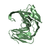
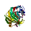
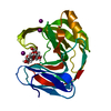
- Structure visualization
| Structure viewer | Molecule: MolmilJmol/JSmol |
|---|
- Downloads & links
-Download
| PDBx/mmCIF format | 1bk1.cif.gz | 45.8 KB | Display | PDBx/mmCIF format |
|---|---|---|---|---|
| PDB format | pdb1bk1.ent.gz | 35 KB | Display | PDB format |
| PDBx/mmJSON format | 1bk1.json.gz | Tree view | PDBx/mmJSON format | |
| Others |  Other downloads Other downloads |
-Validation report
| Arichive directory | https://data.pdbj.org/pub/pdb/validation_reports/bk/1bk1ftp://data.pdbj.org/pub/pdb/validation_reports/bk/1bk1 | HTTPS FTP |
|---|
-Related structure data
| Related structure data |  1xynS S: Starting model for refinement |
|---|---|
| Similar structure data |


-Links
 PDBj
PDBj
- Assembly
| Deposited unit | 
| ||||||||
|---|---|---|---|---|---|---|---|---|---|
| 1 |
| ||||||||
| Unit cell |
|
-Components
| #1: Protein | Mass: 19893.988 Da / Num. of mol.: 1 Source method: isolated from a genetically manipulated source Source: (gene. exp.) Aspergillus kawachii (mold) / Strain: IFO 4308 / Cellular location: EXTRACELLULARGlossary of biology / Gene: XYNC / Plasmid: PUAMXC1 / Cellular location (production host): EXTRACELLULAR / Gene (production host): XYNC / Production host: Aspergillus kawachii (mold) / Strain (production host): IFO 4308 / References: UniProt: P33557, endo-1,4-beta-xylanase |
|---|---|
| #2: Water | ChemComp-HOH / Water Mass: 18.015 Da / Num. of mol.: 111 / Source method: isolated from a natural source / Formula: H2O Mass: 18.015 Da / Num. of mol.: 111 / Source method: isolated from a natural source / Formula: H2O |
-Experimental details
-Experiment
| Experiment | Method: X-RAY DIFFRACTION / Number of used crystals: 2 |
|---|
- Sample preparation
| Crystal | Density Matthews: 2.75 Å3/Da / Density % sol: 55.2 % Description: DATA WERE COLLECTED USING THE WEISSENBERG METHOD | |||||||||||||||||||||||||
|---|---|---|---|---|---|---|---|---|---|---|---|---|---|---|---|---|---|---|---|---|---|---|---|---|---|---|
| Crystal grow | pH: 6.5 / Details: pH 6.5 | |||||||||||||||||||||||||
| Crystal grow | *PLUS Temperature: 25 ℃ / pH: 3.5 / Method: vapor diffusion, hanging dropDetails: 0.02ml of protein solution was mixed with 0.01ml of reservoir solution | |||||||||||||||||||||||||
| Components of the solutions | *PLUS
|
-Data collection
| Diffraction | Mean temperature: 293 K |
|---|---|
| Diffraction source | Source: SYNCHROTRON / Site: Photon Factory  / Beamline: BL-6A / Wavelength: 1 / Beamline: BL-6A / Wavelength: 1 |
| Detector | Type: FUJI / Detector: IMAGE PLATE / Date: Jul 4, 1996 |
| Radiation | Monochromatic (M) / Laue (L): M / Scattering type: x-ray |
| Radiation wavelength | Wavelength: 1 Å / Relative weight: 1 |
| Reflection | Resolution: 2→34.7 Å / Num. obs: 15428 / % possible obs: 98.4 % / Observed criterion σ(I): 0.1 / Redundancy: 8.7 % / Biso Wilson estimate: 27.9 Å2 / Rmerge(I) obs: 0.073 / Net I/σ(I): 10.3 |
| Reflection shell | Resolution: 2→2.07 Å / Rmerge(I) obs: 0.397 / % possible all: 97.2 |
| Reflection | *PLUS Num. measured all: 134544 |
| Reflection shell | *PLUS % possible obs: 97.2 % |
- Processing
| Software |
| ||||||||||||||||||||||||||||||||||||||||||||||||||||||||||||
|---|---|---|---|---|---|---|---|---|---|---|---|---|---|---|---|---|---|---|---|---|---|---|---|---|---|---|---|---|---|---|---|---|---|---|---|---|---|---|---|---|---|---|---|---|---|---|---|---|---|---|---|---|---|---|---|---|---|---|---|---|---|
| Refinement | Method to determine structure: MOLECULAR REPLACEMENT Starting model: TRICHODERMA REESEI XYNI, PDB ENTRY 1XYN Resolution: 2→6 Å / Rfactor Rfree error: 0.01 / Cross valid method: THROUGHOUT / σ(F): 3
| ||||||||||||||||||||||||||||||||||||||||||||||||||||||||||||
| Displacement parameters | Biso mean: 30.1 Å2 | ||||||||||||||||||||||||||||||||||||||||||||||||||||||||||||
| Refine analyze | Luzzati coordinate error obs: 0.27 Å | ||||||||||||||||||||||||||||||||||||||||||||||||||||||||||||
| Refinement step | Cycle: LAST / Resolution: 2→6 Å
| ||||||||||||||||||||||||||||||||||||||||||||||||||||||||||||
| Refine LS restraints |
| ||||||||||||||||||||||||||||||||||||||||||||||||||||||||||||
| LS refinement shell | Resolution: 2→2.09 Å / Rfactor Rfree error: 0.032 / Total num. of bins used: 8
| ||||||||||||||||||||||||||||||||||||||||||||||||||||||||||||
| Xplor file |
| ||||||||||||||||||||||||||||||||||||||||||||||||||||||||||||
| Software | *PLUS Name: X-PLOR / Version: 3.1 / Classification: refinement | ||||||||||||||||||||||||||||||||||||||||||||||||||||||||||||
| Refinement | *PLUS Rfactor Rfree: 0.259 | ||||||||||||||||||||||||||||||||||||||||||||||||||||||||||||
| Solvent computation | *PLUS | ||||||||||||||||||||||||||||||||||||||||||||||||||||||||||||
| Displacement parameters | *PLUS | ||||||||||||||||||||||||||||||||||||||||||||||||||||||||||||
| Refine LS restraints | *PLUS
| ||||||||||||||||||||||||||||||||||||||||||||||||||||||||||||
| LS refinement shell | *PLUS Rfactor obs: 0.309 |
