 Movie
Movie Controller


+ Open data

- Basic information
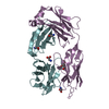
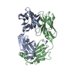
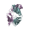
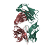
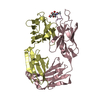
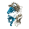
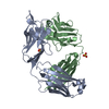
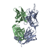
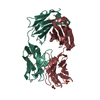
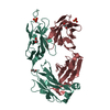
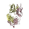
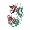
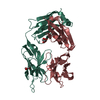
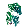
| Entry | Database: PDB / ID: 1bey | ||||||
|---|---|---|---|---|---|---|---|
| Title | ANTIBODY TO CAMPATH-1H HUMANIZED FAB | ||||||
 Components Components | (CAMPATH-1H ANTIBODY) x 2 | ||||||
 Keywords Keywords |  ANTIBODY / FAB / CAMPATH-1H / CD52 ANTIBODY / FAB / CAMPATH-1H / CD52 | ||||||
| Function / homology |  Function and homology information Function and homology information | ||||||
| Biological species |  Homo sapiens (human) Homo sapiens (human) | ||||||
| Method | X-RAY DIFFRACTION / SYNCHROTRON / MOLECULAR REPLACEMENT / Resolution: 3.25 Å | ||||||
 Authors Authors | Cheetham, G.M.T. / Hale, G. / Waldmann, H. / Bloomer, A.C. | ||||||
 Citation Citation | Journal: J.Mol.Biol. / Year: 1998 Title: Crystal structures of a rat anti-CD52 (CAMPATH-1) therapeutic antibody Fab fragment and its humanized counterpart. Authors: Cheetham, G.M. / Hale, G. / Waldmann, H. / Bloomer, A.C. | ||||||
| History |
|
- Structure visualization
| Structure viewer | Molecule: MolmilJmol/JSmol |
|---|
- Downloads & links
-Download
| PDBx/mmCIF format | 1bey.cif.gz | 82.6 KB | Display | PDBx/mmCIF format |
|---|---|---|---|---|
| PDB format | pdb1bey.ent.gz | 66.1 KB | Display | PDB format |
| PDBx/mmJSON format | 1bey.json.gz | Tree view | PDBx/mmJSON format | |
| Others |  Other downloads Other downloads |
-Validation report
| Arichive directory | https://data.pdbj.org/pub/pdb/validation_reports/be/1beyftp://data.pdbj.org/pub/pdb/validation_reports/be/1bey | HTTPS FTP |
|---|
-Related structure data
| Related structure data |  1bfoC 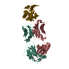 1fdlS  7fabS S: Starting model for refinement C: citing same article ( |
|---|---|
| Similar structure data |


-Links
 PDBj
PDBj

- Assembly
| Deposited unit | 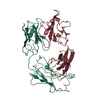
| ||||||||
|---|---|---|---|---|---|---|---|---|---|
| 1 |
| ||||||||
| Unit cell |
|
-Components
| #1: Antibody | Mass: 23596.271 Da / Num. of mol.: 1 / Fragment: FAB FRAGMENT / Source method: isolated from a natural source / Details: HYBRIDOMA CULTURE SUPERNATANT / Source: (natural) Homo sapiens (human) / References: GenBank: 243868, UniProt: Q6GMX0*PLUS |
|---|---|
| #2: Antibody | Mass: 23418.307 Da / Num. of mol.: 1 / Fragment: FAB FRAGMENT / Source method: isolated from a natural source / Details: HYBRIDOMA CULTURE SUPERNATANT / Source: (natural) Homo sapiens (human) / References: GenBank: 243866, UniProt: Q6GMX6*PLUS |
-Experimental details
-Experiment
| Experiment | Method: X-RAY DIFFRACTION / Number of used crystals: 2 |
|---|
- Sample preparation
| Crystal | Density Matthews: 3.4 Å3/Da / Density % sol: 58 % | ||||||||||||||||||||
|---|---|---|---|---|---|---|---|---|---|---|---|---|---|---|---|---|---|---|---|---|---|
| Crystal grow | pH: 7 / Details: pH 7.0 | ||||||||||||||||||||
| Crystal grow | *PLUS Method: vapor diffusion, sitting drop | ||||||||||||||||||||
| Components of the solutions | *PLUS
|
-Data collection
| Diffraction | Mean temperature: 293 K |
|---|---|
| Diffraction source | Source: SYNCHROTRON / Site: SRS  / Beamline: PX9.5 / Wavelength: 0.9 / Beamline: PX9.5 / Wavelength: 0.9 |
| Detector | Type: MARRESEARCH / Detector: IMAGE PLATE / Date: Dec 1, 1992 / Details: MIRRORS |
| Radiation | Monochromator: MONOCHROMATOR / Monochromatic (M) / Laue (L): M / Scattering type: x-ray |
| Radiation wavelength | Wavelength: 0.9 Å / Relative weight: 1 |
| Reflection | Resolution: 3.25→20 Å / Num. obs: 10139 / % possible obs: 92 % / Observed criterion σ(I): 1 / Redundancy: 3 % / Biso Wilson estimate: 39 Å2 / Rmerge(I) obs: 0.11 / Rsym value: 0.11 / Net I/σ(I): 8 |
| Reflection shell | Resolution: 3.25→3.42 Å / Redundancy: 1.6 % / Rmerge(I) obs: 0.204 / Mean I/σ(I) obs: 3.8 / Rsym value: 0.204 / % possible all: 67 |
| Reflection shell | *PLUS % possible obs: 86.6 % / Rmerge(I) obs: 0.227 |
- Processing
| Software |
| ||||||||||||||||||||||||||||||||||||||||||||||||||||||||||||
|---|---|---|---|---|---|---|---|---|---|---|---|---|---|---|---|---|---|---|---|---|---|---|---|---|---|---|---|---|---|---|---|---|---|---|---|---|---|---|---|---|---|---|---|---|---|---|---|---|---|---|---|---|---|---|---|---|---|---|---|---|---|
| Refinement | Method to determine structure: MOLECULAR REPLACEMENT Starting model: PDB ENTRIES 7FAB (CON) 1FDL (VAR) Resolution: 3.25→6 Å / Rfactor Rfree error: 0.014 / Isotropic thermal model: GROUPED, UNRESTRAINED / Cross valid method: THROUGHOUT / σ(F): 3
| ||||||||||||||||||||||||||||||||||||||||||||||||||||||||||||
| Displacement parameters | Biso mean: 54.8 Å2 | ||||||||||||||||||||||||||||||||||||||||||||||||||||||||||||
| Refine analyze |
| ||||||||||||||||||||||||||||||||||||||||||||||||||||||||||||
| Refinement step | Cycle: LAST / Resolution: 3.25→6 Å
| ||||||||||||||||||||||||||||||||||||||||||||||||||||||||||||
| Refine LS restraints |
| ||||||||||||||||||||||||||||||||||||||||||||||||||||||||||||
| LS refinement shell | Resolution: 3.25→3.35 Å / Rfactor Rfree error: 0.059 / Total num. of bins used: 10
| ||||||||||||||||||||||||||||||||||||||||||||||||||||||||||||
| Xplor file | Serial no: 1 / Param file: CNS_TOPPAR:PROTEIN_REP.PA / Topol file: CNS_TOPPAR:PROTEIN.TOP | ||||||||||||||||||||||||||||||||||||||||||||||||||||||||||||
| Software | *PLUS Name: 'CNS' / Classification: refinement | ||||||||||||||||||||||||||||||||||||||||||||||||||||||||||||
| Refinement | *PLUS Rfactor obs: 0.229 / Rfactor Rfree: 0.309 | ||||||||||||||||||||||||||||||||||||||||||||||||||||||||||||
| Solvent computation | *PLUS | ||||||||||||||||||||||||||||||||||||||||||||||||||||||||||||
| Displacement parameters | *PLUS | ||||||||||||||||||||||||||||||||||||||||||||||||||||||||||||
| Refine LS restraints | *PLUS
| ||||||||||||||||||||||||||||||||||||||||||||||||||||||||||||
| LS refinement shell | *PLUS Rfactor obs: 0.321 |
