 Movie
Movie Controller

[English] 日本語
 Yorodumi
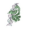
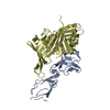
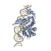
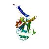
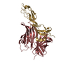
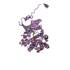
- PDB-1b8x: GLUTATHIONE S-TRANSFERASE FUSED WITH THE NUCLEAR MATRIX TARGETING... -
+ Open data

- Basic information
| Entry | Database: PDB / ID: 1b8x | ||||||
|---|---|---|---|---|---|---|---|
| Title | GLUTATHIONE S-TRANSFERASE FUSED WITH THE NUCLEAR MATRIX TARGETING SIGNAL OF THE TRANSCRIPTION FACTOR AML-1 | ||||||
 Components Components | PROTEIN (AML-1B) | ||||||
 Keywords Keywords |  SIGNAL PROTEIN / NUCLEAR MATRIX TARGETING SIGNAL PROTEIN SIGNAL PROTEIN / NUCLEAR MATRIX TARGETING SIGNAL PROTEIN | ||||||
| Function / homology |  Function and homology information Function and homology information | ||||||
| Biological species |  Escherichia coli (E. coli) Escherichia coli (E. coli) | ||||||
| Method | X-RAY DIFFRACTION / MOLECULAR REPLACEMENT / Resolution: 2.7 Å | ||||||
 Authors Authors | Tang, L. / Guo, B. / Van Wijnen, A.J. / Lian, J.B. / Stein, J.L. / Stein, G.S. / Zhou, G.W. | ||||||
 Citation Citation | Journal: J.Struct.Biol. / Year: 1998 Title: Preliminary crystallographic study of glutathione S-transferase fused with the nuclear matrix targeting signal of the transcription factor AML-1/CBF-alpha2. Authors: Tang, L. / Guo, B. / van Wijnen, A.J. / Lian, J.B. / Stein, J.L. / Stein, G.S. / Zhou, G.W. | ||||||
| History |
|
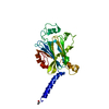
- Structure visualization
| Structure viewer | Molecule: MolmilJmol/JSmol |
|---|
- Downloads & links
-Download
| PDBx/mmCIF format | 1b8x.cif.gz | 62.5 KB | Display | PDBx/mmCIF format |
|---|---|---|---|---|
| PDB format | pdb1b8x.ent.gz | 45.9 KB | Display | PDB format |
| PDBx/mmJSON format | 1b8x.json.gz | Tree view | PDBx/mmJSON format | |
| Others |  Other downloads Other downloads |
-Validation report
| Arichive directory | https://data.pdbj.org/pub/pdb/validation_reports/b8/1b8xftp://data.pdbj.org/pub/pdb/validation_reports/b8/1b8x | HTTPS FTP |
|---|
-Related structure data
| Related structure data | 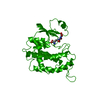 1gneS S: Starting model for refinement |
|---|---|
| Similar structure data |

-Links
 PDBj
PDBj

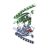
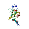
- Assembly
| Deposited unit | 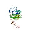
| ||||||||||
|---|---|---|---|---|---|---|---|---|---|---|---|
| 1 |
| ||||||||||
| 2 | 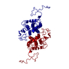
| ||||||||||
| Unit cell |
|
-Components
| #1: Protein | Mass: 32124.119 Da / Num. of mol.: 1 / Fragment: NMTS FRAGMENT Source method: isolated from a genetically manipulated source Source: (gene. exp.) Escherichia coli (E. coli) / Cell line: BL21 / Production host: Escherichia coli (E. coli) / References: UniProt: P08515 |
|---|
-Experimental details
-Experiment
| Experiment | Method: X-RAY DIFFRACTION / Number of used crystals: 1 |
|---|
- Sample preparation
| Crystal | Density Matthews: 2.05 Å3/Da / Density % sol: 50 % | ||||||||||||||||||||||||||||||
|---|---|---|---|---|---|---|---|---|---|---|---|---|---|---|---|---|---|---|---|---|---|---|---|---|---|---|---|---|---|---|---|
| Crystal grow | pH: 7.5 / Details: pH 7.5 | ||||||||||||||||||||||||||||||
| Components of the solutions |
| ||||||||||||||||||||||||||||||
| Crystal grow | *PLUS Method: vapor diffusion, hanging dropDetails: drop consists of equal volume of protein and reservoir solutions | ||||||||||||||||||||||||||||||
| Components of the solutions | *PLUS
|
-Data collection
| Diffraction | Mean temperature: 85 K |
|---|---|
| Diffraction source | Source: ROTATING ANODE / Type: RIGAKU RU300 / Wavelength: 1.5418 |
| Detector | Type: MARRESEARCH / Detector: IMAGE PLATE / Details: MIRRORS |
| Radiation | Monochromator: NI FILTER / Protocol: SINGLE WAVELENGTH / Monochromatic (M) / Laue (L): M / Scattering type: x-ray |
| Radiation wavelength | Wavelength: 1.5418 Å / Relative weight: 1 |
| Reflection | Resolution: 2.7→15 Å / Num. obs: 7449 / % possible obs: 94.9 % / Observed criterion σ(I): 2 / Redundancy: 2.5 % / Biso Wilson estimate: 41 Å2 / Rsym value: 11 / Net I/σ(I): 3 |
| Reflection shell | Resolution: 2.7→2.82 Å / Redundancy: 2 % / Mean I/σ(I) obs: 2.5 / Rsym value: 25 / % possible all: 87.5 |
| Reflection | *PLUS Rmerge(I) obs: 0.11 |
| Reflection shell | *PLUS % possible obs: 87.5 % / Rmerge(I) obs: 0.25 |
- Processing
| Software |
| ||||||||||||||||||||||||||||||||||||||||||||||||||||||||||||||||||||||||||||||||
|---|---|---|---|---|---|---|---|---|---|---|---|---|---|---|---|---|---|---|---|---|---|---|---|---|---|---|---|---|---|---|---|---|---|---|---|---|---|---|---|---|---|---|---|---|---|---|---|---|---|---|---|---|---|---|---|---|---|---|---|---|---|---|---|---|---|---|---|---|---|---|---|---|---|---|---|---|---|---|---|---|---|
| Refinement | Method to determine structure: MOLECULAR REPLACEMENT Starting model: 1GNE Resolution: 2.7→6 Å / Isotropic thermal model: RESTRAINED / Cross valid method: THROUGHOUT / σ(F): 2
| ||||||||||||||||||||||||||||||||||||||||||||||||||||||||||||||||||||||||||||||||
| Refinement step | Cycle: LAST / Resolution: 2.7→6 Å
| ||||||||||||||||||||||||||||||||||||||||||||||||||||||||||||||||||||||||||||||||
| Refine LS restraints |
| ||||||||||||||||||||||||||||||||||||||||||||||||||||||||||||||||||||||||||||||||
| LS refinement shell | Resolution: 2.7→2.81 Å / Total num. of bins used: 8
| ||||||||||||||||||||||||||||||||||||||||||||||||||||||||||||||||||||||||||||||||
| Xplor file |
| ||||||||||||||||||||||||||||||||||||||||||||||||||||||||||||||||||||||||||||||||
| Software | *PLUS Name: X-PLOR / Version: 3.851 / Classification: refinement | ||||||||||||||||||||||||||||||||||||||||||||||||||||||||||||||||||||||||||||||||
| Refinement | *PLUS Rfactor obs: 0.209 | ||||||||||||||||||||||||||||||||||||||||||||||||||||||||||||||||||||||||||||||||
| Solvent computation | *PLUS | ||||||||||||||||||||||||||||||||||||||||||||||||||||||||||||||||||||||||||||||||
| Displacement parameters | *PLUS | ||||||||||||||||||||||||||||||||||||||||||||||||||||||||||||||||||||||||||||||||
| LS refinement shell | *PLUS Rfactor obs: 0.27 |
