 Movie
Movie Controller


+ Open data

- Basic information
| Entry | Database: PDB / ID: 1b3t | ||||||
|---|---|---|---|---|---|---|---|
| Title | EBNA-1 NUCLEAR PROTEIN/DNA COMPLEX | ||||||
 Components Components |
| ||||||
 Keywords Keywords | PROTEIN/DNA /  NUCLEAR PROTEIN / PROTEIN-DNA COMPLEX / DNA-BINDING / ACTIVATOR / ORIGIN-BINDING PROTEIN NUCLEAR PROTEIN / PROTEIN-DNA COMPLEX / DNA-BINDING / ACTIVATOR / ORIGIN-BINDING PROTEIN | ||||||
| Function / homology |  Function and homology information Function and homology informationhost cell PML body / symbiont-mediated suppression of host antigen processing and presentation / viral latency / Hydrolases; Acting on ester bonds; Endodeoxyribonucleases producing 5'-phosphomonoesters / enzyme-substrate adaptor activity / symbiont-mediated disruption of host cell PML body / regulation of DNA replication / endonuclease activity / DNA-binding transcription factor activity / virus-mediated perturbation of host defense response ...host cell PML body / symbiont-mediated suppression of host antigen processing and presentation / viral latency / Hydrolases; Acting on ester bonds; Endodeoxyribonucleases producing 5'-phosphomonoesters / enzyme-substrate adaptor activity / symbiont-mediated disruption of host cell PML body / regulation of DNA replication / endonuclease activity / DNA-binding transcription factor activity / virus-mediated perturbation of host defense response / host cell nucleus / positive regulation of DNA-templated transcription / DNA binding / nucleusSimilarity search - Function | ||||||
| Biological species |  Human herpesvirus 4 (Epstein-Barr virus) Human herpesvirus 4 (Epstein-Barr virus) | ||||||
| Method | X-RAY DIFFRACTION / SYNCHROTRON / MOLECULAR REPLACEMENT / Resolution: 2.2 Å | ||||||
 Authors Authors | Bochkarev, A. / Bochkareva, E. / Edwards, A. / Frappier, L. | ||||||
 Citation Citation | Journal: J.Mol.Biol. / Year: 1998 Title: The 2.2 A structure of a permanganate-sensitive DNA site bound by the Epstein-Barr virus origin binding protein, EBNA1. Authors: Bochkarev, A. / Bochkareva, E. / Frappier, L. / Edwards, A.M. #1: Journal: Cell(Cambridge,Mass.) / Year: 1996Title: Crystal Structure of the DNA-Binding Domain of Theepstein-Barr Virus Origin- Binding Protein, EBNA1, Boundto DNA Authors: Bochkarev, A. / Barwell, J.A. / Pfuetzner, R.A. / Bochkareva, E. / Frappier, L. / Edwards, A.M. #2: Journal: J.Biol.Chem. / Year: 1995Title: Overexpression, Purification, and Crystallization of the DNA Binding and Dimerization Domains of the Epstein-Barr Virus Nuclear Antigen 1 Authors: Barwell, J.A. / Bochkarev, A. / Pfuetzner, R.A. / Tong, H. / Yang, D.S. / Frappier, L. / Edwards, A.M. | ||||||
| History |
|
- Structure visualization
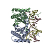
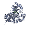
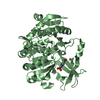
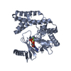
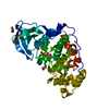
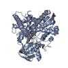
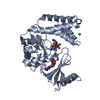
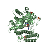
| Structure viewer | Molecule: MolmilJmol/JSmol |
|---|
- Downloads & links
-Download
| PDBx/mmCIF format | 1b3t.cif.gz | 93.2 KB | Display | PDBx/mmCIF format |
|---|---|---|---|---|
| PDB format | pdb1b3t.ent.gz | 66.6 KB | Display | PDB format |
| PDBx/mmJSON format | 1b3t.json.gz | Tree view | PDBx/mmJSON format | |
| Others |  Other downloads Other downloads |
-Validation report
| Arichive directory | https://data.pdbj.org/pub/pdb/validation_reports/b3/1b3tftp://data.pdbj.org/pub/pdb/validation_reports/b3/1b3t | HTTPS FTP |
|---|
-Related structure data
| Related structure data | 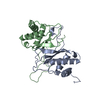 1vhiS S: Starting model for refinement |
|---|---|
| Similar structure data |


-Links
 PDBj
PDBj


- Assembly
| Deposited unit | 
| ||||||||||
|---|---|---|---|---|---|---|---|---|---|---|---|
| 1 |
| ||||||||||
| Unit cell |
|
-Components
| #1: DNA chain | Mass: 5516.578 Da / Num. of mol.: 2 / Source method: obtained synthetically #2: Protein | Mass: 16008.498 Da / Num. of mol.: 2 Fragment: DNA-BINDING AND DIMERIZATION DOMAIN RESIDUES 459 - 607 Source method: isolated from a genetically manipulated source Source: (gene. exp.) Human herpesvirus 4 (Epstein-Barr virus)Genus: Lymphocryptovirus / Strain: GD1 / Cellular location: NUCLEAR / Plasmid: PET15B / Production host:  Escherichia coli (E. coli) / Strain (production host): BL21(DE3)PLUSS / References: UniProt: Q69477, UniProt: P03211*PLUS Escherichia coli (E. coli) / Strain (production host): BL21(DE3)PLUSS / References: UniProt: Q69477, UniProt: P03211*PLUS#3: Water | ChemComp-HOH / | Water Mass: 18.015 Da / Num. of mol.: 182 / Source method: isolated from a natural source / Formula: H2O Mass: 18.015 Da / Num. of mol.: 182 / Source method: isolated from a natural source / Formula: H2OCompound details | EBNA1 ENCODED BY EBV B95-8 COMPRISES 641 AMINO ACIDS AND IS VERY STABLE DIMER BOTH IN SOLUTION AND ...EBNA1 ENCODED BY EBV B95-8 COMPRISES 641 AMINO ACIDS AND IS VERY STABLE DIMER BOTH IN SOLUTION AND WHEN BOUND TO 1 8 BP RECOGNITIO | |
|---|
-Experimental details
-Experiment
| Experiment | Method: X-RAY DIFFRACTION / Number of used crystals: 1 |
|---|
- Sample preparation
| Crystal | Density Matthews: 2.52 Å3/Da / Density % sol: 51.17 % | ||||||||||||||||||||||||||||||||||||||||||||||||||||||||||||||||||||||||||||||||||||
|---|---|---|---|---|---|---|---|---|---|---|---|---|---|---|---|---|---|---|---|---|---|---|---|---|---|---|---|---|---|---|---|---|---|---|---|---|---|---|---|---|---|---|---|---|---|---|---|---|---|---|---|---|---|---|---|---|---|---|---|---|---|---|---|---|---|---|---|---|---|---|---|---|---|---|---|---|---|---|---|---|---|---|---|---|---|
| Crystal grow | Temperature: 277 K / Method: vapor diffusion, hanging drop / pH: 5.6 Details: METHOD - HANGING DROP VAPOR DIFFUSION. RESERVOIR: 100 MM MES (PH 5.6) 20% PEG 4000, 500 MM NACL, 10 MM MGCL2, 10 MM DTT PROTEIN: 10MG/ML 1MM HEPES PH 7.2 1M NACL, 10 MM DTT DRY DNA ...Details: METHOD - HANGING DROP VAPOR DIFFUSION. RESERVOIR: 100 MM MES (PH 5.6) 20% PEG 4000, 500 MM NACL, 10 MM MGCL2, 10 MM DTT PROTEIN: 10MG/ML 1MM HEPES PH 7.2 1M NACL, 10 MM DTT DRY DNA RESUSPENDED IN THE PROTEIN CONTAINING SOLUTION IN MOLAR RATIO 1.5 (DSDNA) / 1.0 (EBNA1 DIMER) DROP: 50% PROTEIN/DNA SOLUTION 50% RESERVOIR SOLUTION CRYSTALLIZATION TEMPERATURE: 4 GEDREES C, vapor diffusion - hanging drop, temperature 277K PH range: 5.6-7.2 | ||||||||||||||||||||||||||||||||||||||||||||||||||||||||||||||||||||||||||||||||||||
| Components of the solutions |
| ||||||||||||||||||||||||||||||||||||||||||||||||||||||||||||||||||||||||||||||||||||
| Crystal grow | *PLUS pH: 7.2 | ||||||||||||||||||||||||||||||||||||||||||||||||||||||||||||||||||||||||||||||||||||
| Components of the solutions | *PLUS
|
-Data collection
| Diffraction | Mean temperature: 100 K |
|---|---|
| Diffraction source | Source: SYNCHROTRON / Site: CHESS  / Beamline: F1 / Wavelength: 0.922 / Beamline: F1 / Wavelength: 0.922 |
| Detector | Type: CUSTOM-MADE / Detector: CCD / Date: May 1, 1994 |
| Radiation | Protocol: SINGLE WAVELENGTH / Monochromatic (M) / Laue (L): M / Scattering type: x-ray |
| Radiation wavelength | Wavelength: 0.922 Å / Relative weight: 1 |
| Reflection | Resolution: 2.1→20 Å / Num. obs: 25865 / % possible obs: 98.9 % / Observed criterion σ(I): -3 / Redundancy: 5.5 % / Rmerge(I) obs: 0.073 / Net I/σ(I): 4.5 |
| Reflection shell | Resolution: 2.1→2.18 Å / Rmerge(I) obs: 0.121 / Mean I/σ(I) obs: 3.1 / % possible all: 93.2 |
| Reflection | *PLUS Num. measured all: 142136 |
| Reflection shell | *PLUS % possible obs: 74.5 % |
- Processing
| Software |
| ||||||||||||||||||||||||||||||||||||||||||||||||||||||||||||
|---|---|---|---|---|---|---|---|---|---|---|---|---|---|---|---|---|---|---|---|---|---|---|---|---|---|---|---|---|---|---|---|---|---|---|---|---|---|---|---|---|---|---|---|---|---|---|---|---|---|---|---|---|---|---|---|---|---|---|---|---|---|
| Refinement | Method to determine structure: MOLECULAR REPLACEMENT Starting model: 1VHI Resolution: 2.2→6 Å / Data cutoff high absF: 1000000 / Data cutoff low absF: 0.001 / Isotropic thermal model: RESTRAINED / Cross valid method: THROUGHOUT / σ(F): 2
| ||||||||||||||||||||||||||||||||||||||||||||||||||||||||||||
| Refinement step | Cycle: LAST / Resolution: 2.2→6 Å
| ||||||||||||||||||||||||||||||||||||||||||||||||||||||||||||
| Refine LS restraints |
| ||||||||||||||||||||||||||||||||||||||||||||||||||||||||||||
| LS refinement shell | Resolution: 2.2→2.29 Å / Total num. of bins used: 8
| ||||||||||||||||||||||||||||||||||||||||||||||||||||||||||||
| Xplor file |
| ||||||||||||||||||||||||||||||||||||||||||||||||||||||||||||
| Software | *PLUS Name: X-PLOR / Version: 3.843 / Classification: refinement | ||||||||||||||||||||||||||||||||||||||||||||||||||||||||||||
| Refinement | *PLUS Highest resolution: 2.2 Å / Lowest resolution: 6 Å / σ(F): 2 / % reflection Rfree: 10 % | ||||||||||||||||||||||||||||||||||||||||||||||||||||||||||||
| Solvent computation | *PLUS | ||||||||||||||||||||||||||||||||||||||||||||||||||||||||||||
| Displacement parameters | *PLUS | ||||||||||||||||||||||||||||||||||||||||||||||||||||||||||||
| Refine LS restraints | *PLUS
| ||||||||||||||||||||||||||||||||||||||||||||||||||||||||||||
| LS refinement shell | *PLUS Rfactor Rfree: 0.274 / % reflection Rfree: 10.1 % / Rfactor Rwork: 0.239 |
