 Movie
Movie Controller

[English] 日本語
 Yorodumi
- PDB-1vhi: EPSTEIN BARR VIRUS NUCLEAR ANTIGEN-1 DNA-BINDING DOMAIN, RESIDUES... -
+ Open data

- Basic information
| Entry | Database: PDB / ID: 1vhi | ||||||
|---|---|---|---|---|---|---|---|
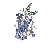
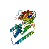
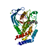
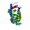
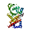
| Title | EPSTEIN BARR VIRUS NUCLEAR ANTIGEN-1 DNA-BINDING DOMAIN, RESIDUES 470-607 | ||||||
 Components Components | EPSTEIN BARR VIRUS NUCLEAR ANTIGEN-1 | ||||||
 Keywords Keywords |  NUCLEAR PROTEIN / DNA-BINDING / ACTIVATOR / ORIGIN-BINDING PROTEIN NUCLEAR PROTEIN / DNA-BINDING / ACTIVATOR / ORIGIN-BINDING PROTEIN | ||||||
| Function / homology |  Function and homology information Function and homology informationhost cell PML body / symbiont-mediated suppression of host antigen processing and presentation / viral latency / Hydrolases; Acting on ester bonds; Endodeoxyribonucleases producing 5'-phosphomonoesters / enzyme-substrate adaptor activity / symbiont-mediated disruption of host cell PML body / regulation of DNA replication / endonuclease activity / DNA-binding transcription factor activity / virus-mediated perturbation of host defense response ...host cell PML body / symbiont-mediated suppression of host antigen processing and presentation / viral latency / Hydrolases; Acting on ester bonds; Endodeoxyribonucleases producing 5'-phosphomonoesters / enzyme-substrate adaptor activity / symbiont-mediated disruption of host cell PML body / regulation of DNA replication / endonuclease activity / DNA-binding transcription factor activity / virus-mediated perturbation of host defense response / positive regulation of DNA-templated transcription / DNA bindingSimilarity search - Function | ||||||
| Biological species |  Human herpesvirus 4 (Epstein-Barr virus) Human herpesvirus 4 (Epstein-Barr virus) | ||||||
| Method | X-RAY DIFFRACTION / MIRAS / Resolution: 2.5 Å | ||||||
 Authors Authors | Bochkarev, A. / Barwell, J. / Pfuetzner, R. / Furey, W. / Edwards, A. / Frappier, L. | ||||||
 Citation Citation | Journal: Cell(Cambridge,Mass.) / Year: 1995 Title: Crystal structure of the DNA-binding domain of the Epstein-Barr virus origin-binding protein EBNA 1. Authors: Bochkarev, A. / Barwell, J.A. / Pfuetzner, R.A. / Furey Jr., W. / Edwards, A.M. / Frappier, L. #1: Journal: J.Biol.Chem. / Year: 1995Title: Overexpression, Purification, and Crystallization of the DNA Binding and Dimerization Domains of the Epstein-Barr Virus Nuclear Antigen 1 Authors: Barwell, J.A. / Bochkarev, A. / Pfuetzner, R.A. / Tong, H. / Yang, D.S. / Frappier, L. / Edwards, A.M. | ||||||
| History |
|
- Structure visualization
| Structure viewer | Molecule: MolmilJmol/JSmol |
|---|
- Downloads & links
-Download
| PDBx/mmCIF format | 1vhi.cif.gz | 76 KB | Display | PDBx/mmCIF format |
|---|---|---|---|---|
| PDB format | pdb1vhi.ent.gz | 57.7 KB | Display | PDB format |
| PDBx/mmJSON format | 1vhi.json.gz | Tree view | PDBx/mmJSON format | |
| Others |  Other downloads Other downloads |
-Validation report
| Arichive directory | https://data.pdbj.org/pub/pdb/validation_reports/vh/1vhiftp://data.pdbj.org/pub/pdb/validation_reports/vh/1vhi | HTTPS FTP |
|---|
-Related structure data
| Similar structure data |
|---|

-Links
 PDBj
PDBj- Assembly
| Deposited unit | 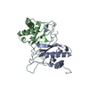
| ||||||||
|---|---|---|---|---|---|---|---|---|---|
| 1 |
| ||||||||
| Unit cell |
|
-Components
| #1: Protein | Mass: 15363.729 Da / Num. of mol.: 2 Fragment: DNA-BINDING AND DIMERIZATION DOMAIN RESIDUES 470 - 607 Source method: isolated from a genetically manipulated source Source: (gene. exp.) Human herpesvirus 4 (Epstein-Barr virus)Genus: Lymphocryptovirus / Strain: GD1 / Gene: E2 / Plasmid: PET15B / Production host:  Escherichia coli (E. coli) / References: UniProt: P03211 Escherichia coli (E. coli) / References: UniProt: P03211#2: Water | ChemComp-HOH / | Water Mass: 18.015 Da / Num. of mol.: 71 / Source method: isolated from a natural source / Formula: H2O Mass: 18.015 Da / Num. of mol.: 71 / Source method: isolated from a natural source / Formula: H2O |
|---|
-Experimental details
-Experiment
| Experiment | Method: X-RAY DIFFRACTION / Number of used crystals: 1 |
|---|
- Sample preparation
| Crystal | Density Matthews: 2.37 Å3/Da / Density % sol: 40 % | ||||||||||||||||||||||||||||||||||||||||||||||||
|---|---|---|---|---|---|---|---|---|---|---|---|---|---|---|---|---|---|---|---|---|---|---|---|---|---|---|---|---|---|---|---|---|---|---|---|---|---|---|---|---|---|---|---|---|---|---|---|---|---|
| Crystal grow | Method: vapor diffusion, hanging drop / pH: 6 Details: HANGING DROP VAPOR DIFFUSION, 50 MM MES PH 6.0 AND 100 MM NACL. SEE REFERENCE 1 FOR MORE DETAILS., vapor diffusion - hanging drop | ||||||||||||||||||||||||||||||||||||||||||||||||
| Crystal | *PLUS | ||||||||||||||||||||||||||||||||||||||||||||||||
| Crystal grow | *PLUS pH: 7.2 / Method: vapor diffusion, hanging drop | ||||||||||||||||||||||||||||||||||||||||||||||||
| Components of the solutions | *PLUS
|
-Data collection
| Diffraction | Mean temperature: 297 K |
|---|---|
| Diffraction source | Source: ROTATING ANODE / Type: RIGAKU RUH2R / Wavelength: 1.5418 |
| Detector | Type: RIGAKU RAXIS IIC / Detector: IMAGE PLATE / Date: Nov 7, 1994 / Details: DOUBLE MIRRORS |
| Radiation | Monochromatic (M) / Laue (L): M / Scattering type: x-ray |
| Radiation wavelength | Wavelength: 1.5418 Å / Relative weight: 1 |
| Reflection | Resolution: 2.5→35 Å / Num. obs: 10110 / % possible obs: 95.9 % / Observed criterion σ(I): 1 / Redundancy: 8.2 % / Rmerge(I) obs: 0.08 / Net I/σ(I): 19.7 |
| Reflection shell | Resolution: 2.5→2.54 Å / Mean I/σ(I) obs: 5.5 / % possible all: 91.3 |
| Reflection | *PLUS Num. measured all: 83324 |
- Processing
| Software |
| ||||||||||||||||||||||||||||||||||||||||||||||||||||||||||||||||||||||||||||||||
|---|---|---|---|---|---|---|---|---|---|---|---|---|---|---|---|---|---|---|---|---|---|---|---|---|---|---|---|---|---|---|---|---|---|---|---|---|---|---|---|---|---|---|---|---|---|---|---|---|---|---|---|---|---|---|---|---|---|---|---|---|---|---|---|---|---|---|---|---|---|---|---|---|---|---|---|---|---|---|---|---|---|
| Refinement | Method to determine structure: MIRAS / Resolution: 2.5→8 Å / σ(F): 2
| ||||||||||||||||||||||||||||||||||||||||||||||||||||||||||||||||||||||||||||||||
| Displacement parameters | Biso mean: 14.4 Å2 | ||||||||||||||||||||||||||||||||||||||||||||||||||||||||||||||||||||||||||||||||
| Refinement step | Cycle: LAST / Resolution: 2.5→8 Å
| ||||||||||||||||||||||||||||||||||||||||||||||||||||||||||||||||||||||||||||||||
| Refine LS restraints |
| ||||||||||||||||||||||||||||||||||||||||||||||||||||||||||||||||||||||||||||||||
| Xplor file |
|
