 Movie
Movie Controller

[English] 日本語
 Yorodumi
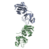
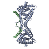
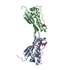
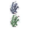
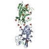
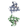
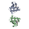
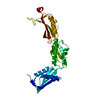
- PDB-1b3j: STRUCTURE OF THE MHC CLASS I HOMOLOG MIC-A, A GAMMADELTA T CELL LIGAND -
+ Open data

- Basic information
| Entry | Database: PDB / ID: 1b3j | |||||||||
|---|---|---|---|---|---|---|---|---|---|---|
| Title | STRUCTURE OF THE MHC CLASS I HOMOLOG MIC-A, A GAMMADELTA T CELL LIGAND | |||||||||
 Components Components | MHC CLASS I HOMOLOG MIC-A | |||||||||
 Keywords Keywords |  IMMUNE SYSTEM / HC I HOMOLOG / HUMAN MICA / MICA / IMMUNOLOGY / MHC / GAMMA-DELTA-TCR / GLYCOPROTEIN / SIGNA IMMUNOGLOBULIN FOLD / T-CELL IMMUNE SYSTEM / HC I HOMOLOG / HUMAN MICA / MICA / IMMUNOLOGY / MHC / GAMMA-DELTA-TCR / GLYCOPROTEIN / SIGNA IMMUNOGLOBULIN FOLD / T-CELL | |||||||||
| Function / homology |  Function and homology information Function and homology informationimmune response to tumor cell / gamma-delta T cell activation / natural killer cell mediated cytotoxicity / natural killer cell lectin-like receptor binding / negative regulation of natural killer cell activation / negative regulation of natural killer cell mediated cytotoxicity / antigen processing and presentation of endogenous peptide antigen via MHC class I via ER pathway, TAP-independent / antigen processing and presentation of endogenous peptide antigen via MHC class Ib / T cell mediated cytotoxicity / positive regulation of T cell mediated cytotoxicity ...immune response to tumor cell / gamma-delta T cell activation / natural killer cell mediated cytotoxicity / natural killer cell lectin-like receptor binding / negative regulation of natural killer cell activation / negative regulation of natural killer cell mediated cytotoxicity / antigen processing and presentation of endogenous peptide antigen via MHC class I via ER pathway, TAP-independent / antigen processing and presentation of endogenous peptide antigen via MHC class Ib / T cell mediated cytotoxicity / positive regulation of T cell mediated cytotoxicity / peptide antigen binding / Immunoregulatory interactions between a Lymphoid and a non-Lymphoid cell / response to heat / defense response to virus / killing of cells of another organism / defense response to bacterium / immune response / external side of plasma membrane / signaling receptor binding / DNA damage response / cell surface / extracellular space / plasma membrane / cytoplasmSimilarity search - Function | |||||||||
| Biological species |  Homo sapiens (human) Homo sapiens (human) | |||||||||
| Method | X-RAY DIFFRACTION / MIR / Resolution: 3 Å | |||||||||
 Authors Authors | Li, P. / Willie, S. / Bauer, S. / Morris, D. / Spies, T. / Strong, R. | |||||||||
 Citation Citation | Journal: Immunity / Year: 1999 Title: Crystal structure of the MHC class I homolog MIC-A, a gammadelta T cell ligand. Authors: Li, P. / Willie, S.T. / Bauer, S. / Morris, D.L. / Spies, T. / Strong, R.K. #1: Journal: Acta Crystallogr.,Sect.D / Year: 1998Title: Expression, Purification, Crystallization and Cryst Graphic Charatterization of the Human Mhc Class I R Protein Mica Authors: Bauer, S. / Willie, S.T. / Spies, T. / Strong, R.K. | |||||||||
| History |
|
- Structure visualization
| Structure viewer | Molecule: MolmilJmol/JSmol |
|---|
- Downloads & links
-Download
| PDBx/mmCIF format | 1b3j.cif.gz | 63.8 KB | Display | PDBx/mmCIF format |
|---|---|---|---|---|
| PDB format | pdb1b3j.ent.gz | 49 KB | Display | PDB format |
| PDBx/mmJSON format | 1b3j.json.gz | Tree view | PDBx/mmJSON format | |
| Others |  Other downloads Other downloads |
-Validation report
| Arichive directory | https://data.pdbj.org/pub/pdb/validation_reports/b3/1b3jftp://data.pdbj.org/pub/pdb/validation_reports/b3/1b3j | HTTPS FTP |
|---|
-Related structure data
| Similar structure data |
|---|


-Links
 PDBj
PDBj- Assembly
| Deposited unit | 
| ||||||||
|---|---|---|---|---|---|---|---|---|---|
| 1 |
| ||||||||
| Unit cell |
| ||||||||
| Components on special symmetry positions |
|
-Components
| #1: Protein | Mass: 31600.236 Da / Num. of mol.: 1 / Fragment: EXTRACELLULAR DOMAIN, RESIDUES 1 - 274 Source method: isolated from a genetically manipulated source Source: (gene. exp.) Homo sapiens (human) / Cellular location: EXTRACELLULARGlossary of biology / Gene: MICA-001 / Cell line (production host): Hi5 / Gene (production host): MICA-001 / Production host:  Trichoplusia ni (cabbage looper) / References: GenBank: 1708676, UniProt: Q29983*PLUS Trichoplusia ni (cabbage looper) / References: GenBank: 1708676, UniProt: Q29983*PLUS |
|---|---|
| #2: Polysaccharide | 2-acetamido-2-deoxy-beta-D-glucopyranose-(1-4)-2-acetamido-2-deoxy-beta-D-glucopyranose / Mass: 424.401 Da / Num. of mol.: 1 Source method: isolated from a genetically manipulated source |
| #3: Water | ChemComp-HOH / Water Mass: 18.015 Da / Num. of mol.: 52 / Source method: isolated from a natural source / Formula: H2O Mass: 18.015 Da / Num. of mol.: 52 / Source method: isolated from a natural source / Formula: H2O |
-Experimental details
-Experiment
| Experiment | Method: X-RAY DIFFRACTION / Number of used crystals: 1 |
|---|
- Sample preparation
| Crystal | Density Matthews: 5.3 Å3/Da / Density % sol: 80 % | ||||||||||||||||||||
|---|---|---|---|---|---|---|---|---|---|---|---|---|---|---|---|---|---|---|---|---|---|
| Crystal grow | pH: 5.5 / Details: pH 5.50 | ||||||||||||||||||||
| Components of the solutions |
| ||||||||||||||||||||
| Crystal grow | *PLUS Temperature: 291 K / Method: vapor diffusion, hanging dropDetails: drop consists of equal volume of protein and reservoir solutions | ||||||||||||||||||||
| Components of the solutions | *PLUS
|
-Data collection
| Diffraction | Mean temperature: 100 K |
|---|---|
| Diffraction source | Source: ROTATING ANODE / Type: RIGAKU RU200 / Wavelength: 1.5418 |
| Detector | Type: RIGAKU RAXIS VI / Detector: IMAGE PLATE / Date: May 1, 1998 / Details: YALE MIRRORS |
| Radiation | Protocol: SINGLE WAVELENGTH / Monochromatic (M) / Laue (L): M / Scattering type: x-ray |
| Radiation wavelength | Wavelength: 1.5418 Å / Relative weight: 1 |
| Reflection | Resolution: 3→25 Å / Num. obs: 15536 / % possible obs: 97.9 % / Redundancy: 4 % / Biso Wilson estimate: 70.1 Å2 / Rmerge(I) obs: 0.061 / Rsym value: 0.061 / Net I/σ(I): 23.5 |
| Reflection shell | Resolution: 3→3.05 Å / Redundancy: 3.2 % / Rmerge(I) obs: 0.356 / Mean I/σ(I) obs: 3.2 / Rsym value: 0.356 / % possible all: 96.6 |
| Reflection | *PLUS Highest resolution: 2.8 Å / Num. obs: 19368 / % possible obs: 99.7 % / Num. measured all: 130489 / Rmerge(I) obs: 0.063 |
| Reflection shell | *PLUS % possible obs: 100 % / Rmerge(I) obs: 0.329 / Mean I/σ(I) obs: 5.8 |
- Processing
| Software |
| ||||||||||||||||||||||||||||||||||||||||||||||||||||||||||||
|---|---|---|---|---|---|---|---|---|---|---|---|---|---|---|---|---|---|---|---|---|---|---|---|---|---|---|---|---|---|---|---|---|---|---|---|---|---|---|---|---|---|---|---|---|---|---|---|---|---|---|---|---|---|---|---|---|---|---|---|---|---|
| Refinement | Method to determine structure: MIR / Resolution: 3→15 Å / Rfactor Rfree error: 0.008 / Data cutoff high absF: 10000000 / Data cutoff low absF: 0.001 / Isotropic thermal model: RESTRAINED / Cross valid method: THROUGHOUT / σ(F): 0 Details: BULK SOLVENT MODEL USED BULK SOLVENT MODELING. METHOD USED: FLAT MODEL KSOL: 0.30 BSOL: 50.0 (A**2)
| ||||||||||||||||||||||||||||||||||||||||||||||||||||||||||||
| Solvent computation | Bsol: 50 Å2 / ksol: 0.3 e/Å3 | ||||||||||||||||||||||||||||||||||||||||||||||||||||||||||||
| Displacement parameters | Biso mean: 46.1 Å2 | ||||||||||||||||||||||||||||||||||||||||||||||||||||||||||||
| Refine analyze |
| ||||||||||||||||||||||||||||||||||||||||||||||||||||||||||||
| Refinement step | Cycle: LAST / Resolution: 3→15 Å
| ||||||||||||||||||||||||||||||||||||||||||||||||||||||||||||
| Refine LS restraints |
| ||||||||||||||||||||||||||||||||||||||||||||||||||||||||||||
| LS refinement shell | Resolution: 3→3.19 Å / Rfactor Rfree error: 0.026 / Total num. of bins used: 6
| ||||||||||||||||||||||||||||||||||||||||||||||||||||||||||||
| Xplor file |
| ||||||||||||||||||||||||||||||||||||||||||||||||||||||||||||
| Software | *PLUS Name: X-PLOR / Version: 3.851 / Classification: refinement | ||||||||||||||||||||||||||||||||||||||||||||||||||||||||||||
| Refinement | *PLUS Highest resolution: 2.8 Å / Lowest resolution: 9999 Å / Num. reflection obs: 18416 / Rfactor obs: 0.253 / Rfactor Rfree: 0.293 | ||||||||||||||||||||||||||||||||||||||||||||||||||||||||||||
| Solvent computation | *PLUS | ||||||||||||||||||||||||||||||||||||||||||||||||||||||||||||
| Displacement parameters | *PLUS | ||||||||||||||||||||||||||||||||||||||||||||||||||||||||||||
| Refine LS restraints | *PLUS
| ||||||||||||||||||||||||||||||||||||||||||||||||||||||||||||
| LS refinement shell | *PLUS |
