 Movie
Movie Controller

[English] 日本語
 Yorodumi
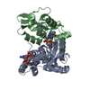
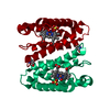
- PDB-1aoh: SINGLE COHESIN DOMAIN FROM THE SCAFFOLDING PROTEIN CIPA OF THE CL... -
+ Open data

- Basic information
| Entry | Database: PDB / ID: 1aoh | ||||||
|---|---|---|---|---|---|---|---|
| Title | SINGLE COHESIN DOMAIN FROM THE SCAFFOLDING PROTEIN CIPA OF THE CLOSTRIDIUM THERMOCELLUM CELLULOSOME | ||||||
 Components Components | Cellulosomal-scaffolding protein A | ||||||
 Keywords Keywords |  STRUCTURAL PROTEIN / CELLULOSOME SUBUNIT / B-BARREL / CELLULOSE DEGRADATION STRUCTURAL PROTEIN / CELLULOSOME SUBUNIT / B-BARREL / CELLULOSE DEGRADATION | ||||||
| Function / homology |  Function and homology informationcellulose binding / cellulose catabolic process / hydrolase activity, hydrolyzing O-glycosyl compounds / cell wall organization / extracellular region Function and homology informationcellulose binding / cellulose catabolic process / hydrolase activity, hydrolyzing O-glycosyl compounds / cell wall organization / extracellular regionSimilarity search - Function | ||||||
| Biological species |  Clostridium thermocellum (bacteria) Clostridium thermocellum (bacteria) | ||||||
| Method | X-RAY DIFFRACTION / SYNCHROTRON / MIR / Resolution: 1.7 Å | ||||||
 Authors Authors | Alzari, P.M. / Tavares, G. | ||||||
 Citation Citation | Journal: J.Mol.Biol. / Year: 1997 Title: The crystal structure of a type I cohesin domain at 1.7 A resolution. Authors: Tavares, G.A. / Beguin, P. / Alzari, P.M. #1: Journal: Protein Sci. / Year: 1996Title: Subcloning of a DNA Fragment Encoding a Single Cohesin Domain of the Clostridium Thermocellum Cellulosome-Integrating Protein Cipa: Purification, Crystallization, and Preliminary Diffraction ...Title: Subcloning of a DNA Fragment Encoding a Single Cohesin Domain of the Clostridium Thermocellum Cellulosome-Integrating Protein Cipa: Purification, Crystallization, and Preliminary Diffraction Analysis of the Encoded Polypeptide Authors: Beguin, P. / Raynaud, O. / Chaveroche, M.K. / Dridi, A. / Alzari, P.M. | ||||||
| History |
|
- Structure visualization
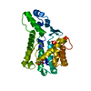
| Structure viewer | Molecule: MolmilJmol/JSmol |
|---|
- Downloads & links
-Download
| PDBx/mmCIF format | 1aoh.cif.gz | 72.5 KB | Display | PDBx/mmCIF format |
|---|---|---|---|---|
| PDB format | pdb1aoh.ent.gz | 54.6 KB | Display | PDB format |
| PDBx/mmJSON format | 1aoh.json.gz | Tree view | PDBx/mmJSON format | |
| Others |  Other downloads Other downloads |
-Validation report
| Arichive directory | https://data.pdbj.org/pub/pdb/validation_reports/ao/1aohftp://data.pdbj.org/pub/pdb/validation_reports/ao/1aoh | HTTPS FTP |
|---|
-Related structure data
| Similar structure data |
|---|


-Links
 PDBj
PDBj
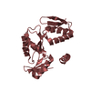
- Assembly
| Deposited unit | 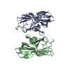
| ||||||||
|---|---|---|---|---|---|---|---|---|---|
| 1 |
| ||||||||
| Unit cell |
| ||||||||
| Noncrystallographic symmetry (NCS) | NCS oper: (Code: given Matrix: (-0.99976, 0.0207, -0.0073), Vector : |
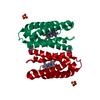
-Components
| #1: Protein | Mass: 15760.738 Da / Num. of mol.: 2 / Fragment: COHESIN DOMAIN residues 1216-1361 Source method: isolated from a genetically manipulated source Source: (gene. exp.) Clostridium thermocellum (strain ATCC 27405 / DSM 1237 / NBRC 103400 / NCIMB 10682 / NRRL B-4536 / VPI 7372) (bacteria)Strain: ATCC 27405 / DSM 1237 / NBRC 103400 / NCIMB 10682 / NRRL B-4536 / VPI 7372 Cell line: BL21 / Cellular location: CYTOPLASM / Gene: cipA, Cthe_3077 / Plasmid: PREP4 / Cell line (production host): BL21 / Cellular location (production host): CYTOPLASM / Gene (production host): LACI / Production host: Escherichia coli (E. coli) / Strain (production host): 293 / References: UniProt: Q06851#2: Water | ChemComp-HOH / | Water Mass: 18.015 Da / Num. of mol.: 381 / Source method: isolated from a natural source / Formula: H2O Mass: 18.015 Da / Num. of mol.: 381 / Source method: isolated from a natural source / Formula: H2OCompound details | THE WILD TYPE PROTEIN CIPA CONTAINS NINE HOMOLOGOUS COHESIN DOMAINS (THE STRUCTURE PRESENTED HERE ...THE WILD TYPE PROTEIN CIPA CONTAINS NINE HOMOLOGOUS | |
|---|
-Experimental details
-Experiment
| Experiment | Method: X-RAY DIFFRACTION / Number of used crystals: 1 |
|---|
- Sample preparation
| Crystal | Density Matthews: 2.1 Å3/Da / Density % sol: 40 % | ||||||||||||||||||||||||||||||||||||||||||||||||||||||||||||
|---|---|---|---|---|---|---|---|---|---|---|---|---|---|---|---|---|---|---|---|---|---|---|---|---|---|---|---|---|---|---|---|---|---|---|---|---|---|---|---|---|---|---|---|---|---|---|---|---|---|---|---|---|---|---|---|---|---|---|---|---|---|
| Crystal grow | pH: 6.25 Details: PROTEIN WAS CRYSTALLIZED FROM 18% PEG-8000, 0.2 M CALCIUM ACETATE, 6% GLYCEROL AND 0.05 M SODIUM CACODYLATE, PH 6.25 | ||||||||||||||||||||||||||||||||||||||||||||||||||||||||||||
| Crystal | *PLUS | ||||||||||||||||||||||||||||||||||||||||||||||||||||||||||||
| Crystal grow | *PLUS Method: vapor diffusion, hanging drop / Details: Beguin, P., (1996) Protein Sci., 5, 1192 | ||||||||||||||||||||||||||||||||||||||||||||||||||||||||||||
| Components of the solutions | *PLUS
|
-Data collection
| Diffraction | Mean temperature: 110 K |
|---|---|
| Diffraction source | Source: SYNCHROTRON / Site: LURE  / Beamline: DW32 / Wavelength: 0.983 / Beamline: DW32 / Wavelength: 0.983 |
| Detector | Type: MARRESEARCH / Detector: IMAGE PLATE / Date: Nov 1, 1996 |
| Radiation | Monochromatic (M) / Laue (L): M / Scattering type: x-ray |
| Radiation wavelength | Wavelength: 0.983 Å / Relative weight: 1 |
| Reflection | Resolution: 1.7→20 Å / Num. obs: 29669 / % possible obs: 92.2 % / Redundancy: 5.8 % / Biso Wilson estimate: 14 Å2 / Rmerge(I) obs: 0.055 / Net I/σ(I): 22.2 |
| Reflection shell | Resolution: 1.7→1.76 Å / Redundancy: 3.2 % / Rmerge(I) obs: 0.253 / Mean I/σ(I) obs: 4.5 / % possible all: 86.6 |
| Reflection | *PLUS Num. measured all: 175259 |
| Reflection shell | *PLUS % possible obs: 86.6 % |
- Processing
| Software |
| ||||||||||||||||||||||||||||||||||||||||||||||||||||||||||||
|---|---|---|---|---|---|---|---|---|---|---|---|---|---|---|---|---|---|---|---|---|---|---|---|---|---|---|---|---|---|---|---|---|---|---|---|---|---|---|---|---|---|---|---|---|---|---|---|---|---|---|---|---|---|---|---|---|---|---|---|---|---|
| Refinement | Method to determine structure: MIR / Resolution: 1.7→10 Å / Rfactor Rfree error: 0.007 / Data cutoff high absF: 0 / Data cutoff low absF: 0 / Cross valid method: THROUGHOUT / σ(F): 0
| ||||||||||||||||||||||||||||||||||||||||||||||||||||||||||||
| Refinement step | Cycle: LAST / Resolution: 1.7→10 Å
| ||||||||||||||||||||||||||||||||||||||||||||||||||||||||||||
| Refine LS restraints |
| ||||||||||||||||||||||||||||||||||||||||||||||||||||||||||||
| Refine LS restraints NCS | NCS model details: UNRESTRAINED | ||||||||||||||||||||||||||||||||||||||||||||||||||||||||||||
| LS refinement shell | Resolution: 1.7→1.78 Å / Total num. of bins used: 8
| ||||||||||||||||||||||||||||||||||||||||||||||||||||||||||||
| Xplor file |
| ||||||||||||||||||||||||||||||||||||||||||||||||||||||||||||
| Software | *PLUS Name: X-PLOR / Version: 3.1 / Classification: refinement | ||||||||||||||||||||||||||||||||||||||||||||||||||||||||||||
| Refinement | *PLUS Rfactor Rfree: 0.26 | ||||||||||||||||||||||||||||||||||||||||||||||||||||||||||||
| Solvent computation | *PLUS | ||||||||||||||||||||||||||||||||||||||||||||||||||||||||||||
| Displacement parameters | *PLUS | ||||||||||||||||||||||||||||||||||||||||||||||||||||||||||||
| Refine LS restraints | *PLUS
| ||||||||||||||||||||||||||||||||||||||||||||||||||||||||||||
| LS refinement shell | *PLUS Rfactor Rwork: 0.29 |
